5VA9

Human pancreatic alpha amylase in complex with peptide inhibitor piHA-L5(d10Y)


Experimental Data Snapshot

  • Method: X-RAY DIFFRACTION
  • Resolution: 2.55 Å
  • R-Value Free: 0.230 
  • R-Value Work: 0.196 
  • R-Value Observed: 0.198 

wwPDB Validation   3D Report Full Report


This is version 2.0 of the entry. See complete history


Literature

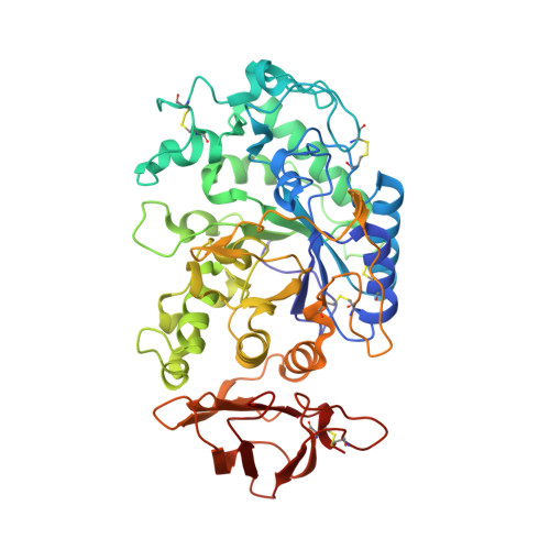
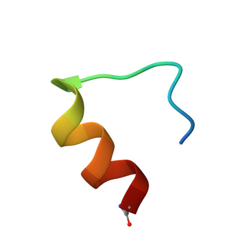
Folding Then Binding vs Folding Through Binding in Macrocyclic Peptide Inhibitors of Human Pancreatic alpha-Amylase.

Goldbach, L.Vermeulen, B.J.A.Caner, S.Liu, M.Tysoe, C.van Gijzel, L.Yoshisada, R.Trellet, M.van Ingen, H.Brayer, G.D.Bonvin, A.M.J.J.Jongkees, S.A.K.

(2019) ACS Chem Biol 14: 1751-1759

  • DOI: https://doi.org/10.1021/acschembio.9b00290
  • Primary Citation of Related Structures:  
    5U3A, 5VA9

  • PubMed Abstract: 

    De novo macrocyclic peptides, derived using selection technologies such as phage and mRNA display, present unique and unexpected solutions to challenging biological problems. This is due in part to their unusual folds, which are able to present side chains in ways not available to canonical structures such as α-helices and β-sheets. Despite much recent interest in these molecules, their folding and binding behavior remains poorly characterized. In this work, we present cocrystallization, docking, and solution NMR structures of three de novo macrocyclic peptides that all bind as competitive inhibitors with single-digit nanomolar K i to the active site of human pancreatic α-amylase. We show that a short stably folded motif in one of these is nucleated by internal hydrophobic interactions in an otherwise dynamic conformation in solution. Comparison of the solution structures with a target-bound structure from docking indicates that stabilization of the bound conformation is provided through interactions with the target protein after binding. These three structures also reveal a surprising functional convergence to present a motif of a single arginine sandwiched between two aromatic residues in the interactions of the peptide with the key catalytic residues of the enzyme, despite little to no other structural homology. Our results suggest that intramolecular hydrophobic interactions are important for priming binding of small macrocyclic peptides to their target and that high rigidity is not necessary for high affinity.


  • Organizational Affiliation

    Department of Chemical Biology and Drug Discovery , Utrecht Institute of Pharmaceutical Sciences, Utrecht University , 3584 CG Utrecht , The Netherlands.


Macromolecules
Find similar proteins by:  (by identity cutoff)  |  3D Structure
Entity ID: 1
MoleculeChains Sequence LengthOrganismDetailsImage
Pancreatic alpha-amylase
A, B
496Homo sapiensMutation(s): 0 
Gene Names: AMY2A
EC: 3.2.1.1
UniProt & NIH Common Fund Data Resources
Find proteins for P04746 (Homo sapiens)
Explore P04746 
Go to UniProtKB:  P04746
PHAROS:  P04746
GTEx:  ENSG00000243480 
Entity Groups  
Sequence Clusters30% Identity50% Identity70% Identity90% Identity95% Identity100% Identity
UniProt GroupP04746
Sequence Annotations
Expand
  • Reference Sequence

Find similar proteins by:  Sequence   |   3D Structure  

Entity ID: 2
MoleculeChains Sequence LengthOrganismDetailsImage
Peptide Inhibitor piHA-L5(d10Y)
C, D
20synthetic constructMutation(s): 0 
Entity Groups  
Sequence Clusters30% Identity50% Identity70% Identity90% Identity95% Identity100% Identity
Sequence Annotations
Expand
  • Reference Sequence
Experimental Data & Validation

Experimental Data

  • Method: X-RAY DIFFRACTION
  • Resolution: 2.55 Å
  • R-Value Free: 0.230 
  • R-Value Work: 0.196 
  • R-Value Observed: 0.198 
  • Space Group: P 43 21 2
Unit Cell:
Length ( Å )Angle ( ˚ )
a = 169.92α = 90
b = 169.92β = 90
c = 79.42γ = 90
Software Package:
Software NamePurpose
PHENIXrefinement
XDSdata reduction
XSCALEdata scaling
PHASERphasing

Structure Validation

View Full Validation Report


Entry History & Funding Information

Deposition Data


Funding OrganizationLocationGrant Number
GlycoNet: National Centres of ExcellenceCanadaDO-2

Revision History  (Full details and data files)

  • Version 1.0: 2018-03-28
    Type: Initial release
  • Version 1.1: 2018-04-18
    Changes: Data collection
  • Version 1.2: 2019-09-25
    Changes: Data collection, Database references
  • Version 2.0: 2023-10-04
    Changes: Data collection, Database references, Derived calculations, Polymer sequence, Refinement description