5V3F

Co-crystal structure of the fluorogenic RNA Mango


Experimental Data Snapshot

  • Method: X-RAY DIFFRACTION
  • Resolution: 1.70 Å
  • R-Value Free: 0.203 
  • R-Value Work: 0.196 
  • R-Value Observed: 0.197 

wwPDB Validation   3D Report Full Report


Ligand Structure Quality Assessment 


This is version 1.3 of the entry. See complete history


Literature

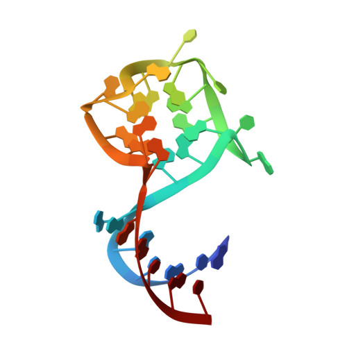
Structural basis for high-affinity fluorophore binding and activation by RNA Mango.

Trachman, R.J.Demeshkina, N.A.Lau, M.W.L.Panchapakesan, S.S.S.Jeng, S.C.Y.Unrau, P.J.Ferre-D'Amare, A.R.

(2017) Nat Chem Biol 13: 807-813

  • DOI: https://doi.org/10.1038/nchembio.2392
  • Primary Citation of Related Structures:  
    5V3F

  • PubMed Abstract: 

    Genetically encoded fluorescent protein tags have revolutionized proteome studies, whereas the lack of intrinsically fluorescent RNAs has hindered transcriptome exploration. Among several RNA-fluorophore complexes that potentially address this problem, RNA Mango has an exceptionally high affinity for its thiazole orange (TO)-derived fluorophore, TO1-Biotin (K d ∼3 nM), and, in complex with related ligands, it is one of the most redshifted fluorescent macromolecular tags known. To elucidate how this small aptamer exhibits such properties, which make it well suited for studying low-copy cellular RNAs, we determined its 1.7-Å-resolution co-crystal structure. Unexpectedly, the entire ligand, including TO, biotin and the linker connecting them, abuts one of the near-planar faces of the three-tiered G-quadruplex. The two heterocycles of TO are held in place by two loop adenines and form a 45° angle with respect to each other. Minimizing this angle would increase quantum yield and further improve this tool for in vivo RNA visualization.


  • Organizational Affiliation

    Biochemistry and Biophysics Center, National Heart, Lung and Blood Institute, Bethesda, Maryland, USA.


Macromolecules
Find similar nucleic acids by:  (by identity cutoff)  |  3D Structure
Entity ID: 1
MoleculeChains LengthOrganismImage
RNA (31-MER)
A, B
31synthetic construct
Sequence Annotations
Expand
  • Reference Sequence
Small Molecules
Ligands 3 Unique
IDChains Name / Formula / InChI Key2D Diagram3D Interactions
74G
Query on 74G

Download Ideal Coordinates CCD File 
F [auth A],
J [auth B]
4-{[(2S)-3-{2,16-dioxo-20-[(3aS,4S,6aR)-2-oxohexahydro-1H-thieno[3,4-d]imidazol-4-yl]-6,9,12-trioxa-3,15-diazaicosan-1-yl}-2,3-dihydro-1,3-benzothiazol-2-yl]methyl}-1-methylquinolin-1-ium
C38 H51 N6 O6 S2
BXBHTLUUPXOIJA-SDUHKGLLSA-O
PO4
Query on PO4

Download Ideal Coordinates CCD File 
K [auth B],
L [auth B]
PHOSPHATE ION
O4 P
NBIIXXVUZAFLBC-UHFFFAOYSA-K
K
Query on K

Download Ideal Coordinates CCD File 
C [auth A]
D [auth A]
E [auth A]
G [auth B]
H [auth B]
C [auth A],
D [auth A],
E [auth A],
G [auth B],
H [auth B],
I [auth B]
POTASSIUM ION
K
NPYPAHLBTDXSSS-UHFFFAOYSA-N
Experimental Data & Validation

Experimental Data

  • Method: X-RAY DIFFRACTION
  • Resolution: 1.70 Å
  • R-Value Free: 0.203 
  • R-Value Work: 0.196 
  • R-Value Observed: 0.197 
  • Space Group: P 41 21 2
Unit Cell:
Length ( Å )Angle ( ˚ )
a = 63.199α = 90
b = 63.199β = 90
c = 138.413γ = 90
Software Package:
Software NamePurpose
PHENIXrefinement
HKL-2000data scaling
SHELXCDphasing

Structure Validation

View Full Validation Report


Ligand Structure Quality Assessment 


Entry History 

Deposition Data

Revision History  (Full details and data files)

  • Version 1.0: 2017-05-24
    Type: Initial release
  • Version 1.1: 2017-06-14
    Changes: Database references
  • Version 1.2: 2017-06-28
    Changes: Database references
  • Version 1.3: 2024-03-06
    Changes: Data collection, Database references, Derived calculations