5UXM

Type II DAH7PS from Pseudomonas aeruginosa with Trp bound


Experimental Data Snapshot

  • Method: X-RAY DIFFRACTION
  • Resolution: 1.54 Å
  • R-Value Free: 0.210 
  • R-Value Work: 0.185 
  • R-Value Observed: 0.186 

wwPDB Validation   3D Report Full Report


Ligand Structure Quality Assessment 


This is version 1.4 of the entry. See complete history


Literature

A Pseudoisostructural Type II DAH7PS Enzyme from Pseudomonas aeruginosa: Alternative Evolutionary Strategies to Control Shikimate Pathway Flux.

Sterritt, O.W.Kessans, S.A.Jameson, G.B.Parker, E.J.

(2018) Biochemistry 57: 2667-2678

  • DOI: https://doi.org/10.1021/acs.biochem.8b00082
  • Primary Citation of Related Structures:  
    5UXM, 5UXN, 5UXO

  • PubMed Abstract: 

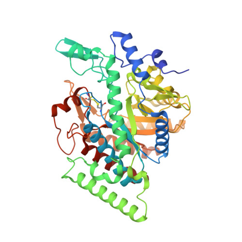
    The shikimate pathway is responsible for the biosynthesis of key aromatic metabolites in microorganisms and plants. The enzyme 3-deoxy-d- arabino-heptulosonate 7-phosphate synthase (DAH7PS) catalyzes the first step of the pathway and DAH7PSs are classified as either type I or type II. The DAH7PSs from Pseudomonas aeruginosa are of particular interest as open reading frames encoding four putative DAH7PS isoenzymes, two classified as type Iα and two classified as type II, have been identified. Here, the structure of a type II DAH7PS enzyme from P. aeruginosa (PAO1) has been determined at 1.54 Å resolution, in complex with its allosteric inhibitor tryptophan. Structural differences in the extra-barrel elements, when compared to other type II DAH7PS enzymes, directly relate to the formation of a distinct quaternary conformation with consequences for allosteric function and the control of flux to branching pathways. In contrast to the well-characterized Mycobacterium tuberculosis type II DAH7PS, which binds multiple allosteric inhibitors, this PaeDAH7PS PA2843 is observed to be modestly allosterically inhibited by a single aromatic amino acid, tryptophan. In addition, structures in complex with tyrosine or with no allosteric ligand bound were determined. These structures provide new insights into the linkages between the active and allosteric sites. With four putative DAH7PS enzymes, P. aeruginosa appears to have evolved control of shikimate pathway flux at the genetic level, rather than control by multiple allosteric effectors to a single type II DAH7PS, as in M. tuberculosis. Type II DAH7PSs, thus, appear to have a more varied evolutionary trajectory than previously indicated.


  • Organizational Affiliation

    Biomolecular Interaction Centre and School of Physical and Chemical Sciences , University of Canterbury , Christchurch 8041 , New Zealand.


Macromolecules
Find similar proteins by:  (by identity cutoff)  |  3D Structure
Entity ID: 1
MoleculeChains Sequence LengthOrganismDetailsImage
Phospho-2-dehydro-3-deoxyheptonate aldolase453Pseudomonas aeruginosa PAO1Mutation(s): 0 
Gene Names: PA2843
EC: 2.5.1.54
UniProt
Find proteins for Q9I000 (Pseudomonas aeruginosa (strain ATCC 15692 / DSM 22644 / CIP 104116 / JCM 14847 / LMG 12228 / 1C / PRS 101 / PAO1))
Explore Q9I000 
Go to UniProtKB:  Q9I000
Entity Groups  
Sequence Clusters30% Identity50% Identity70% Identity90% Identity95% Identity100% Identity
UniProt GroupQ9I000
Sequence Annotations
Expand
  • Reference Sequence
Small Molecules
Ligands 5 Unique
IDChains Name / Formula / InChI Key2D Diagram3D Interactions
TRP
Query on TRP

Download Ideal Coordinates CCD File 
C [auth A]TRYPTOPHAN
C11 H12 N2 O2
QIVBCDIJIAJPQS-VIFPVBQESA-N
PEP
Query on PEP

Download Ideal Coordinates CCD File 
B [auth A]PHOSPHOENOLPYRUVATE
C3 H5 O6 P
DTBNBXWJWCWCIK-UHFFFAOYSA-N
PO4
Query on PO4

Download Ideal Coordinates CCD File 
D [auth A],
E [auth A]
PHOSPHATE ION
O4 P
NBIIXXVUZAFLBC-UHFFFAOYSA-K
CO
Query on CO

Download Ideal Coordinates CCD File 
F [auth A]COBALT (II) ION
Co
XLJKHNWPARRRJB-UHFFFAOYSA-N
CL
Query on CL

Download Ideal Coordinates CCD File 
G [auth A],
H [auth A]
CHLORIDE ION
Cl
VEXZGXHMUGYJMC-UHFFFAOYSA-M
Binding Affinity Annotations 
IDSourceBinding Affinity
TRP Binding MOAD:  5UXM Kd: 3600 (nM) from 1 assay(s)
Experimental Data & Validation

Experimental Data

  • Method: X-RAY DIFFRACTION
  • Resolution: 1.54 Å
  • R-Value Free: 0.210 
  • R-Value Work: 0.185 
  • R-Value Observed: 0.186 
  • Space Group: I 2 2 2
Unit Cell:
Length ( Å )Angle ( ˚ )
a = 81.68α = 90
b = 100.37β = 90
c = 115.6γ = 90
Software Package:
Software NamePurpose
REFMACrefinement
iMOSFLMdata reduction
Aimlessdata scaling
MOLREPphasing

Structure Validation

View Full Validation Report


Ligand Structure Quality Assessment 


Entry History 

Deposition Data

Revision History  (Full details and data files)

  • Version 1.0: 2018-02-28
    Type: Initial release
  • Version 1.1: 2018-05-02
    Changes: Data collection, Database references
  • Version 1.2: 2018-05-16
    Changes: Data collection, Database references
  • Version 1.3: 2019-11-20
    Changes: Derived calculations
  • Version 1.4: 2023-10-04
    Changes: Data collection, Database references, Refinement description