


The crystal structure of chloramphenicol acetyltransferase from Vibrio fischeri ES114
Tan, K., Zhou, M., Anderson, W.F., Joachimiak, A.To be published.
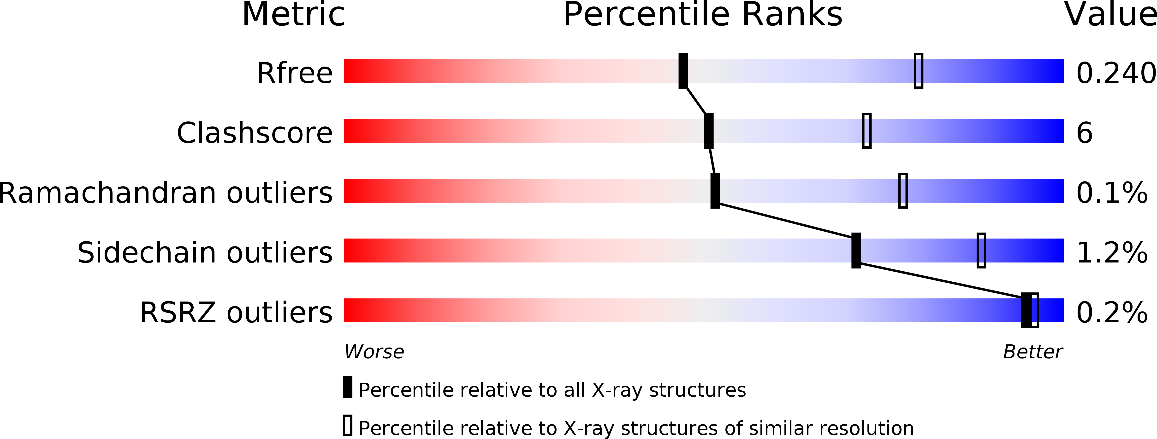
Experimental Data Snapshot
wwPDB Validation 3D Report Full Report
Entity ID: 1 | |||||
|---|---|---|---|---|---|
| Molecule | Chains | Sequence Length | Organism | Details | Image |
| Chloramphenicol acetyltransferase | 221 | Aliivibrio fischeri ES114 | Mutation(s): 0 Gene Names: VF_A0790 EC: 2.3.1.28 | 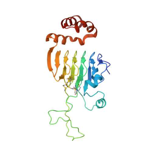 | |
UniProt | |||||
Find proteins for Q5DZD6 (Aliivibrio fischeri (strain ATCC 700601 / ES114)) Explore Q5DZD6 Go to UniProtKB: Q5DZD6 | |||||
Entity Groups | |||||
| Sequence Clusters | 30% Identity50% Identity70% Identity90% Identity95% Identity100% Identity | ||||
| UniProt Group | Q5DZD6 | ||||
Sequence AnnotationsExpand | |||||
| |||||
| Ligands 5 Unique | |||||
|---|---|---|---|---|---|
| ID | Chains | Name / Formula / InChI Key | 2D Diagram | 3D Interactions | |
| MES Query on MES | E [auth A] J [auth B] P [auth C] Q [auth C] V [auth D] | 2-(N-MORPHOLINO)-ETHANESULFONIC ACID C6 H13 N O4 S SXGZJKUKBWWHRA-UHFFFAOYSA-N |  | ||
| ACT Query on ACT | G [auth A] H [auth A] L [auth B] S [auth C] T [auth C] | ACETATE ION C2 H3 O2 QTBSBXVTEAMEQO-UHFFFAOYSA-M |  | ||
| FMT Query on FMT | AA [auth D] | FORMIC ACID C H2 O2 BDAGIHXWWSANSR-UHFFFAOYSA-N |  | ||
| CL Query on CL | I [auth A] M [auth B] N [auth B] O [auth B] U [auth C] | CHLORIDE ION Cl VEXZGXHMUGYJMC-UHFFFAOYSA-M |  | ||
| MG Query on MG | F [auth A], K [auth B], R [auth C], X [auth D] | MAGNESIUM ION Mg JLVVSXFLKOJNIY-UHFFFAOYSA-N |  | ||
| Modified Residues 1 Unique | |||||
|---|---|---|---|---|---|
| ID | Chains | Type | Formula | 2D Diagram | Parent |
| MSE Query on MSE | A, B, C, D | L-PEPTIDE LINKING | C5 H11 N O2 Se |  | MET |
| Length ( Å ) | Angle ( ˚ ) |
|---|---|
| a = 147.426 | α = 90 |
| b = 147.426 | β = 90 |
| c = 101.211 | γ = 120 |
| Software Name | Purpose |
|---|---|
| PHENIX | refinement |
| HKL-3000 | data reduction |
| HKL-3000 | data scaling |
| HKL-3000 | phasing |
| Funding Organization | Location | Grant Number |
|---|---|---|
| National Institutes of Health/National Institute Of Allergy and Infectious Diseases (NIH/NIAID) | United States | HHSN272201200026C |
RCSB PDB (citation) is hosted by
RCSB PDB is a member of the

