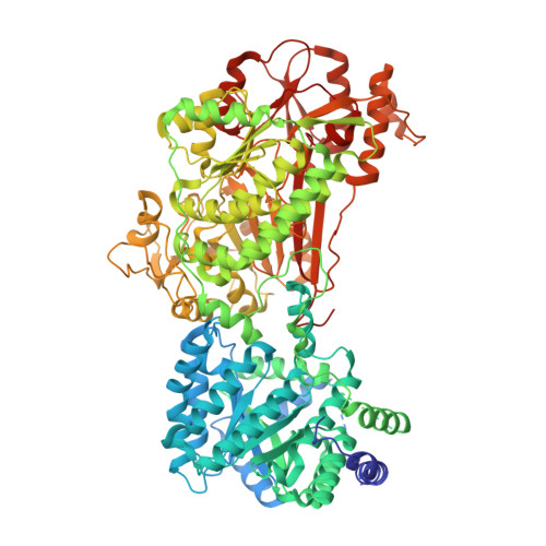
5UX5

Structure of Proline Utilization A (PutA) from Corynebacterium freiburgense


Experimental Data Snapshot

  • Method: X-RAY DIFFRACTION
  • Resolution: 2.70 Å
  • R-Value Free: 0.236 
  • R-Value Work: 0.183 
  • R-Value Observed: 0.185 

wwPDB Validation   3D Report Full Report


Ligand Structure Quality Assessment 


This is version 1.5 of the entry. See complete history


Literature

Structure and characterization of a class 3B proline utilization A: Ligand-induced dimerization and importance of the C-terminal domain for catalysis.

Korasick, D.A.Gamage, T.T.Christgen, S.Stiers, K.M.Beamer, L.J.Henzl, M.T.Becker, D.F.Tanner, J.J.

(2017) J Biol Chem 292: 9652-9665

  • DOI: https://doi.org/10.1074/jbc.M117.786855
  • Primary Citation of Related Structures:  
    5UX5

  • PubMed Abstract: 

    The bifunctional flavoenzyme proline utilization A (PutA) catalyzes the two-step oxidation of proline to glutamate using separate proline dehydrogenase (PRODH) and l-glutamate-γ-semialdehyde dehydrogenase active sites. Because PutAs catalyze sequential reactions, they are good systems for studying how metabolic enzymes communicate via substrate channeling. Although mechanistically similar, PutAs vary widely in domain architecture, oligomeric state, and quaternary structure, and these variations represent different structural solutions to the problem of sequestering a reactive metabolite. Here, we studied PutA from Corynebacterium freiburgense (CfPutA), which belongs to the uncharacterized 3B class of PutAs. A 2.7 Å resolution crystal structure showed the canonical arrangement of PRODH, l-glutamate-γ-semialdehyde dehydrogenase, and C-terminal domains, including an extended interdomain tunnel associated with substrate channeling. The structure unexpectedly revealed a novel open conformation of the PRODH active site, which is interpreted to represent the non-activated conformation, an elusive form of PutA that exhibits suboptimal channeling. Nevertheless, CfPutA exhibited normal substrate-channeling activity, indicating that it isomerizes into the active state under assay conditions. Sedimentation-velocity experiments provided insight into the isomerization process, showing that CfPutA dimerizes in the presence of a proline analog and NAD + These results are consistent with the morpheein model of enzyme hysteresis, in which substrate binding induces conformational changes that promote assembly of a high-activity oligomer. Finally, we used domain deletion analysis to investigate the function of the C-terminal domain. Although this domain contains neither catalytic residues nor substrate sites, its removal impaired both catalytic activities, suggesting that it may be essential for active-site integrity.


  • Organizational Affiliation

    From the Departments of Biochemistry and.


Macromolecules
Find similar proteins by:  (by identity cutoff)  |  3D Structure
Entity ID: 1
MoleculeChains Sequence LengthOrganismDetailsImage
BIFUNCTIONAL PROTEIN Proline utilization A (PutA)
A, B, C, D
1,076Corynebacterium freiburgenseMutation(s): 0 
EC: 1.2.1.88
UniProt
Find proteins for A0A1X8XLF1 (Corynebacterium freiburgense)
Explore A0A1X8XLF1 
Go to UniProtKB:  A0A1X8XLF1
Entity Groups  
Sequence Clusters30% Identity50% Identity70% Identity90% Identity95% Identity100% Identity
UniProt GroupA0A1X8XLF1
Sequence Annotations
Expand
  • Reference Sequence
Small Molecules
Ligands 3 Unique
IDChains Name / Formula / InChI Key2D Diagram3D Interactions
FAD
Query on FAD

Download Ideal Coordinates CCD File 
E [auth A],
H [auth B],
K [auth C],
N [auth D]
FLAVIN-ADENINE DINUCLEOTIDE
C27 H33 N9 O15 P2
VWWQXMAJTJZDQX-UYBVJOGSSA-N
NAD
Query on NAD

Download Ideal Coordinates CCD File 
F [auth A],
I [auth B],
L [auth C],
O [auth D]
NICOTINAMIDE-ADENINE-DINUCLEOTIDE
C21 H27 N7 O14 P2
BAWFJGJZGIEFAR-NNYOXOHSSA-N
SO4
Query on SO4

Download Ideal Coordinates CCD File 
G [auth A],
J [auth B],
M [auth C],
P [auth D]
SULFATE ION
O4 S
QAOWNCQODCNURD-UHFFFAOYSA-L
Experimental Data & Validation

Experimental Data

  • Method: X-RAY DIFFRACTION
  • Resolution: 2.70 Å
  • R-Value Free: 0.236 
  • R-Value Work: 0.183 
  • R-Value Observed: 0.185 
  • Space Group: P 1
Unit Cell:
Length ( Å )Angle ( ˚ )
a = 84.959α = 71.48
b = 116.36β = 89.68
c = 140.734γ = 83.42
Software Package:
Software NamePurpose
PHENIXrefinement
Aimlessdata scaling
PDB_EXTRACTdata extraction
XDSdata reduction
BALBESphasing

Structure Validation

View Full Validation Report


Ligand Structure Quality Assessment 


Entry History & Funding Information

Deposition Data

  • Released Date: 2017-04-26 
  • Deposition Author(s): Tanner, J.J.

Funding OrganizationLocationGrant Number
National Institutes of Health/National Institute of General Medical Sciences (NIH/NIGMS)United StatesR01GM065546
National Institutes of Health/National Institute of General Medical Sciences (NIH/NIGMS)United StatesR01GM061068
National Institutes of Health/National Institute of General Medical Sciences (NIH/NIGMS)United StatesP30GM103335
National Science Foundation (NSF, United States)United StatesDBI-1156692

Revision History  (Full details and data files)

  • Version 1.0: 2017-04-26
    Type: Initial release
  • Version 1.1: 2017-05-03
    Changes: Database references
  • Version 1.2: 2017-06-21
    Changes: Database references
  • Version 1.3: 2017-09-13
    Changes: Author supporting evidence
  • Version 1.4: 2019-11-27
    Changes: Author supporting evidence
  • Version 1.5: 2023-10-04
    Changes: Data collection, Database references, Refinement description