5UWD

Crystal structure of EGFR kinase domain (L858R, T790M, V948R) in complex with the covalent inhibitor CO-1686


Experimental Data Snapshot

  • Method: X-RAY DIFFRACTION
  • Resolution: 3.06 Å
  • R-Value Free: 0.280 
  • R-Value Work: 0.213 
  • R-Value Observed: 0.216 

wwPDB Validation   3D Report Full Report


Ligand Structure Quality Assessment 


This is version 1.1 of the entry. See complete history


Literature

Proteome-wide Map of Targets of T790M-EGFR-Directed Covalent Inhibitors.

Niessen, S.Dix, M.M.Barbas, S.Potter, Z.E.Lu, S.Brodsky, O.Planken, S.Behenna, D.Almaden, C.Gajiwala, K.S.Ryan, K.Ferre, R.Lazear, M.R.Hayward, M.M.Kath, J.C.Cravatt, B.F.

(2017) Cell Chem Biol 24: 1388-1400.e7

  • DOI: https://doi.org/10.1016/j.chembiol.2017.08.017
  • Primary Citation of Related Structures:  
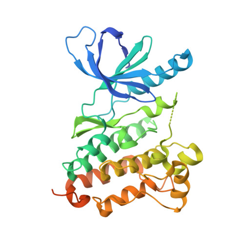
    5UWD

  • PubMed Abstract: 

    Patients with non-small cell lung cancers that have kinase-activating epidermal growth factor receptor (EGFR) mutations are highly responsive to first- and second-generation EGFR inhibitors. However, these patients often relapse due to a secondary, drug-resistant mutation in EGFR whereby the gatekeeper threonine is converted to methionine (T790M). Several third-generation EGFR inhibitors have been developed that irreversibly inactivate T790M-EGFR while sparing wild-type EGFR, thus reducing epithelium-based toxicities. Using chemical proteomics, we show here that individual T790M-EGFR inhibitors exhibit strikingly distinct off-target profiles in human cells. The FDA-approved drug osimertinib (AZD9291), in particular, was found to covalently modify cathepsins in cell and animal models, which correlated with lysosomal accumulation of the drug. Our findings thus show how chemical proteomics can be used to differentiate covalent kinase inhibitors based on global selectivity profiles in living systems and identify specific off-targets of these inhibitors that may affect drug activity and safety.


  • Organizational Affiliation

    Worldwide Medicinal Chemistry, La Jolla Laboratories, Pfizer Worldwide Research and Development, 10770 Science Center Drive, San Diego, CA 92121, USA; Oncology RU, La Jolla Laboratories, Pfizer Worldwide Research and Development, 10770 Science Center Drive, San Diego, CA 92121, USA. Electronic address: sherry.niessen@pfizer.com.


Macromolecules
Find similar proteins by:  (by identity cutoff)  |  3D Structure
Entity ID: 1
MoleculeChains Sequence LengthOrganismDetailsImage
Epidermal growth factor receptor329Homo sapiensMutation(s): 3 
Gene Names: EGFRERBBERBB1HER1
EC: 2.7.10.1
UniProt & NIH Common Fund Data Resources
Find proteins for P00533 (Homo sapiens)
Explore P00533 
Go to UniProtKB:  P00533
PHAROS:  P00533
GTEx:  ENSG00000146648 
Entity Groups  
Sequence Clusters30% Identity50% Identity70% Identity90% Identity95% Identity100% Identity
UniProt GroupP00533
Sequence Annotations
Expand
  • Reference Sequence
Small Molecules
Ligands 1 Unique
IDChains Name / Formula / InChI Key2D Diagram3D Interactions
8OV
Query on 8OV

Download Ideal Coordinates CCD File 
B [auth A]N-(3-{[2-{[4-(4-acetylpiperazin-1-yl)-2-methoxyphenyl]amino}-5-(trifluoromethyl)pyrimidin-4-yl]amino}phenyl)propanamide
C27 H30 F3 N7 O3
XRXFHVJORXTVSS-UHFFFAOYSA-N
Experimental Data & Validation

Experimental Data

  • Method: X-RAY DIFFRACTION
  • Resolution: 3.06 Å
  • R-Value Free: 0.280 
  • R-Value Work: 0.213 
  • R-Value Observed: 0.216 
  • Space Group: P 61 2 2
Unit Cell:
Length ( Å )Angle ( ˚ )
a = 78.926α = 90
b = 78.926β = 90
c = 215.596γ = 120
Software Package:
Software NamePurpose
BUSTERrefinement
XDSdata reduction
SCALAdata scaling
PHASERphasing

Structure Validation

View Full Validation Report


Ligand Structure Quality Assessment 


Entry History 

Deposition Data

Revision History  (Full details and data files)

  • Version 1.0: 2017-10-18
    Type: Initial release
  • Version 1.1: 2017-11-29
    Changes: Database references