5UUO

Crystal structure of SARO_2595 from Novosphingobium aromaticivorans


Experimental Data Snapshot

  • Method: X-RAY DIFFRACTION
  • Resolution: 1.25 Å
  • R-Value Free: 0.132 
  • R-Value Work: 0.130 
  • R-Value Observed: 0.130 

wwPDB Validation   3D Report Full Report


Ligand Structure Quality Assessment 


This is version 2.1 of the entry. See complete history


Literature

Novosphingobium aromaticivoransuses a Nu-class glutathioneS-transferase as a glutathione lyase in breaking the beta-aryl ether bond of lignin.

Kontur, W.S.Bingman, C.A.Olmsted, C.N.Wassarman, D.R.Ulbrich, A.Gall, D.L.Smith, R.W.Yusko, L.M.Fox, B.G.Noguera, D.R.Coon, J.J.Donohue, T.J.

(2018) J Biol Chem 293: 4955-4968

  • DOI: https://doi.org/10.1074/jbc.RA117.001268
  • Primary Citation of Related Structures:  
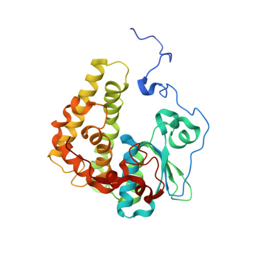
    5UUN, 5UUO

  • PubMed Abstract: 

    As a major component of plant cell walls, lignin is a potential renewable source of valuable chemicals. Several sphingomonad bacteria have been identified that can break the β-aryl ether bond connecting most phenylpropanoid units of the lignin heteropolymer. Here, we tested three sphingomonads predicted to be capable of breaking the β-aryl ether bond of the dimeric aromatic compound guaiacylglycerol-β-guaiacyl ether (GGE) and found that Novosphingobium aromaticivorans metabolizes GGE at one of the fastest rates thus far reported. After the ether bond of racemic GGE is broken by replacement with a thioether bond involving glutathione, the glutathione moiety must be removed from the resulting two stereoisomers of the phenylpropanoid conjugate β-glutathionyl-γ-hydroxypropiovanillone (GS-HPV). We found that the Nu-class glutathione S -transferase NaGST Nu is the only enzyme needed to remove glutathione from both ( R )- and ( S )-GS-HPV in N. aromaticivorans We solved the crystal structure of NaGST Nu and used molecular modeling to propose a mechanism for the glutathione lyase (deglutathionylation) reaction in which an enzyme-stabilized glutathione thiolate attacks the thioether bond of GS-HPV, and the reaction proceeds through an enzyme-stabilized enolate intermediate. Three residues implicated in the proposed mechanism (Thr 51 , Tyr 166 , and Tyr 224 ) were found to be critical for the lyase reaction. We also found that Nu-class GSTs from Sphingobium sp. SYK-6 (which can also break the β-aryl ether bond) and Escherichia coli (which cannot break the β-aryl ether bond) can also cleave ( R )- and ( S )-GS-HPV, suggesting that glutathione lyase activity may be common throughout this widespread but largely uncharacterized class of glutathione S -transferases.


  • Organizational Affiliation

    From the Wisconsin Energy Institute.


Macromolecules
Find similar proteins by:  (by identity cutoff)  |  3D Structure
Entity ID: 1
MoleculeChains Sequence LengthOrganismDetailsImage
Glutathione S-transferase-like proteinA [auth B],
B [auth A]
293Novosphingobium aromaticivorans DSM 12444Mutation(s): 0 
Gene Names: Saro_2595
UniProt
Find proteins for Q2G542 (Novosphingobium aromaticivorans (strain ATCC 700278 / DSM 12444 / CCUG 56034 / CIP 105152 / NBRC 16084 / F199))
Explore Q2G542 
Go to UniProtKB:  Q2G542
Entity Groups  
Sequence Clusters30% Identity50% Identity70% Identity90% Identity95% Identity100% Identity
UniProt GroupQ2G542
Sequence Annotations
Expand
  • Reference Sequence
Small Molecules
Ligands 3 Unique
IDChains Name / Formula / InChI Key2D Diagram3D Interactions
GSH
Query on GSH

Download Ideal Coordinates CCD File 
C [auth B],
D [auth B],
Q [auth A],
R [auth A]
GLUTATHIONE
C10 H17 N3 O6 S
RWSXRVCMGQZWBV-WDSKDSINSA-N
SO4
Query on SO4

Download Ideal Coordinates CCD File 
E [auth B],
F [auth B],
G [auth B],
S [auth A]
SULFATE ION
O4 S
QAOWNCQODCNURD-UHFFFAOYSA-L
EDO
Query on EDO

Download Ideal Coordinates CCD File 
AA [auth A]
BA [auth A]
H [auth B]
I [auth B]
J [auth B]
AA [auth A],
BA [auth A],
H [auth B],
I [auth B],
J [auth B],
K [auth B],
L [auth B],
M [auth B],
N [auth B],
O [auth B],
P [auth B],
T [auth A],
U [auth A],
V [auth A],
W [auth A],
X [auth A],
Y [auth A],
Z [auth A]
1,2-ETHANEDIOL
C2 H6 O2
LYCAIKOWRPUZTN-UHFFFAOYSA-N
Experimental Data & Validation

Experimental Data

  • Method: X-RAY DIFFRACTION
  • Resolution: 1.25 Å
  • R-Value Free: 0.132 
  • R-Value Work: 0.130 
  • R-Value Observed: 0.130 
  • Space Group: P 21 21 21
Unit Cell:
Length ( Å )Angle ( ˚ )
a = 68.81α = 90
b = 70.39β = 90
c = 168.23γ = 90
Software Package:
Software NamePurpose
PHENIXrefinement
XSCALEdata reduction
XDSdata scaling
PHASERphasing

Structure Validation

View Full Validation Report


Ligand Structure Quality Assessment 


Entry History & Funding Information

Deposition Data


Funding OrganizationLocationGrant Number
Department of Energy (DOE, United States)United StatesDE-FC02-07ER64494

Revision History  (Full details and data files)

  • Version 1.0: 2018-02-28
    Type: Initial release
  • Version 1.1: 2018-03-28
    Changes: Data collection, Database references
  • Version 2.0: 2018-04-18
    Changes: Atomic model, Data collection, Database references, Derived calculations, Structure summary
  • Version 2.1: 2022-03-16
    Changes: Author supporting evidence, Database references, Derived calculations, Structure summary