5UQ1

Crystal structure of human Cdk2-Spy1 complex


Experimental Data Snapshot

  • Method: X-RAY DIFFRACTION
  • Resolution: 3.20 Å
  • R-Value Free: 0.261 
  • R-Value Work: 0.205 
  • R-Value Observed: 0.208 

wwPDB Validation   3D Report Full Report


This is version 1.2 of the entry. See complete history


Literature

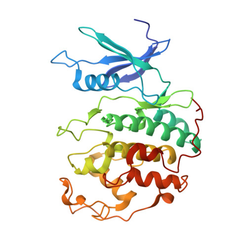
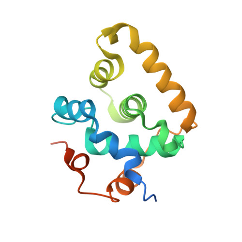
Structural basis of divergent cyclin-dependent kinase activation by Spy1/RINGO proteins.

McGrath, D.A.Fifield, B.A.Marceau, A.H.Tripathi, S.Porter, L.A.Rubin, S.M.

(2017) EMBO J 36: 2251-2262

  • DOI: https://doi.org/10.15252/embj.201796905
  • Primary Citation of Related Structures:  
    5UQ1, 5UQ2, 5UQ3

  • PubMed Abstract: 

    Cyclin-dependent kinases (Cdks) are principal drivers of cell division and are an important therapeutic target to inhibit aberrant proliferation. Cdk enzymatic activity is tightly controlled through cyclin interactions, posttranslational modifications, and binding of inhibitors such as the p27 tumor suppressor protein. Spy1/RINGO (Spy1) proteins bind and activate Cdk but are resistant to canonical regulatory mechanisms that establish cell-cycle checkpoints. Cancer cells exploit Spy1 to stimulate proliferation through inappropriate activation of Cdks, yet the mechanism is unknown. We have determined crystal structures of the Cdk2-Spy1 and p27-Cdk2-Spy1 complexes that reveal how Spy1 activates Cdk. We find that Spy1 confers structural changes to Cdk2 that obviate the requirement of Cdk activation loop phosphorylation. Spy1 lacks the cyclin-binding site that mediates p27 and substrate affinity, explaining why Cdk-Spy1 is poorly inhibited by p27 and lacks specificity for substrates with cyclin-docking sites. We identify mutations in Spy1 that ablate its ability to activate Cdk2 and to proliferate cells. Our structural description of Spy1 provides important mechanistic insights that may be utilized for targeting upregulated Spy1 in cancer.


  • Organizational Affiliation

    Department of Chemistry and Biochemistry, University of California, Santa Cruz, CA, USA.


Macromolecules
Find similar proteins by:  (by identity cutoff)  |  3D Structure
Entity ID: 1
MoleculeChains Sequence LengthOrganismDetailsImage
Cyclin-dependent kinase 2
A, C
301Homo sapiensMutation(s): 0 
Gene Names: CDK2CDKN2
EC: 2.7.11.22
UniProt & NIH Common Fund Data Resources
Find proteins for P24941 (Homo sapiens)
Explore P24941 
Go to UniProtKB:  P24941
PHAROS:  P24941
GTEx:  ENSG00000123374 
Entity Groups  
Sequence Clusters30% Identity50% Identity70% Identity90% Identity95% Identity100% Identity
UniProt GroupP24941
Sequence Annotations
Expand
  • Reference Sequence
Find similar proteins by:  (by identity cutoff)  |  3D Structure
Entity ID: 2
MoleculeChains Sequence LengthOrganismDetailsImage
Speedy protein A
B, D
160Homo sapiensMutation(s): 0 
Gene Names: SPDYASPDY1SPY1
UniProt & NIH Common Fund Data Resources
Find proteins for Q5MJ70 (Homo sapiens)
Explore Q5MJ70 
Go to UniProtKB:  Q5MJ70
PHAROS:  Q5MJ70
GTEx:  ENSG00000163806 
Entity Groups  
Sequence Clusters30% Identity50% Identity70% Identity90% Identity95% Identity100% Identity
UniProt GroupQ5MJ70
Sequence Annotations
Expand
  • Reference Sequence
Experimental Data & Validation

Experimental Data

  • Method: X-RAY DIFFRACTION
  • Resolution: 3.20 Å
  • R-Value Free: 0.261 
  • R-Value Work: 0.205 
  • R-Value Observed: 0.208 
  • Space Group: P 31 2 1
Unit Cell:
Length ( Å )Angle ( ˚ )
a = 75.359α = 90
b = 75.359β = 90
c = 325.791γ = 120
Software Package:
Software NamePurpose
PHENIXrefinement
iMOSFLMdata reduction
Aimlessdata scaling
PHASERphasing

Structure Validation

View Full Validation Report


Entry History 

Deposition Data

Revision History  (Full details and data files)

  • Version 1.0: 2017-07-05
    Type: Initial release
  • Version 1.1: 2017-07-19
    Changes: Database references
  • Version 1.2: 2017-08-09
    Changes: Database references