


Crystal structure of a D-beta-hydroxybutyrate dehydrogenase from Burkholderia multivorans
Mayclin, S.J., Dranow, D.M., Lorimer, D., Edwards, T.E.To be published.
Experimental Data Snapshot
wwPDB Validation 3D Report Full Report

Entity ID: 1 | |||||
|---|---|---|---|---|---|
| Molecule | Chains | Sequence Length | Organism | Details | Image |
| 3-ketoacyl-ACP reductase | 273 | Burkholderia multivorans | Mutation(s): 0 Gene Names: WM33_05200 | 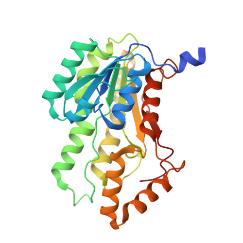 | |
Entity Groups | |||||
| Sequence Clusters | 30% Identity50% Identity70% Identity90% Identity95% Identity100% Identity | ||||
Sequence AnnotationsExpand | |||||
| |||||
| Ligands 2 Unique | |||||
|---|---|---|---|---|---|
| ID | Chains | Name / Formula / InChI Key | 2D Diagram | 3D Interactions | |
| EDO Query on EDO | E [auth A] F [auth A] G [auth A] H [auth A] I [auth A] | 1,2-ETHANEDIOL C2 H6 O2 LYCAIKOWRPUZTN-UHFFFAOYSA-N |  | ||
| NO3 Query on NO3 | J [auth A] | NITRATE ION N O3 NHNBFGGVMKEFGY-UHFFFAOYSA-N |  | ||
| Length ( Å ) | Angle ( ˚ ) |
|---|---|
| a = 64.38 | α = 90 |
| b = 132.09 | β = 108.68 |
| c = 64.53 | γ = 90 |
| Software Name | Purpose |
|---|---|
| XSCALE | data scaling |
| Coot | model building |
| PHENIX | refinement |
| PDB_EXTRACT | data extraction |
| XDS | data reduction |
| MOLREP | phasing |
RCSB PDB (citation) is hosted by
RCSB PDB is a member of the

