5UNJ

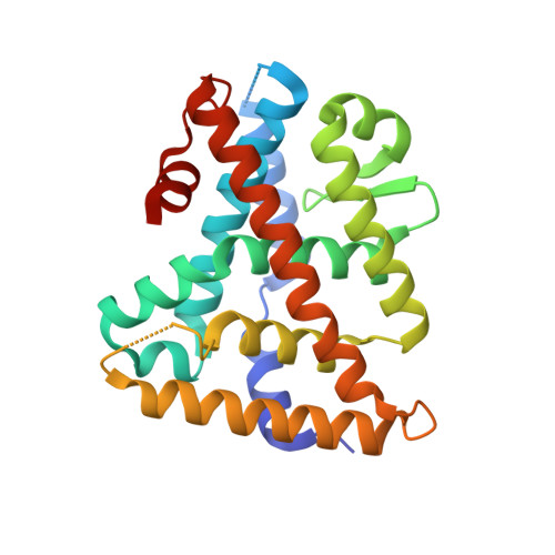
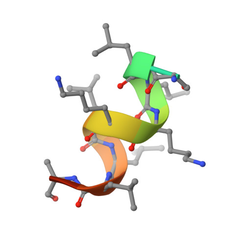
Structure of Human Liver Receptor Homolog 1 in complex with PGC1a and RJW100

  • Classification: NUCLEAR PROTEIN
  • Organism(s): Homo sapiens
  • Expression System: Escherichia coli
  • Mutation(s): No 

  • Deposited: 2017-01-31 Released: 2017-04-19 
  • Deposition Author(s): Mays, S.G., Ortlund, E.A.
  • Funding Organization(s): National Institutes of Health/National Institute of Diabetes and Digestive and Kidney Disease (NIH/NIDDK), National Institutes of Health/National Institute of General Medical Sciences (NIH/NIGMS)

Experimental Data Snapshot

  • Method: X-RAY DIFFRACTION
  • Resolution: 1.96 Å
  • R-Value Free: 0.227 
  • R-Value Work: 0.199 
  • R-Value Observed: 0.201 

wwPDB Validation   3D Report Full Report


Ligand Structure Quality Assessment 


This is version 1.4 of the entry. See complete history


Literature

Structure and Dynamics of the Liver Receptor Homolog 1-PGC1 alpha Complex.

Mays, S.G.Okafor, C.D.Tuntland, M.L.Whitby, R.J.Dharmarajan, V.Stec, J.Griffin, P.R.Ortlund, E.A.

(2017) Mol Pharmacol 92: 1-11

  • DOI: https://doi.org/10.1124/mol.117.108514
  • Primary Citation of Related Structures:  
    5UNJ

  • PubMed Abstract: 

    Peroxisome proliferator-activated gamma coactivator 1- α (PGC1 α ) regulates energy metabolism by directly interacting with transcription factors to modulate gene expression. Among the PGC1 α binding partners is liver receptor homolog 1 (LRH-1; NR5A2), an orphan nuclear hormone receptor that controls lipid and glucose homeostasis. Although PGC1 α is known to bind and activate LRH-1, mechanisms through which PGC1 α changes LRH-1 conformation to drive transcription are unknown. Here, we used biochemical and structural methods to interrogate the LRH-1-PGC1 α complex. Purified, full-length LRH-1, as well as isolated ligand binding domain, bound to PGC1 α with higher affinity than to the coactivator, nuclear receptor coactivator-2 (Tif2), in coregulator peptide recruitment assays. We present the first crystal structure of the LRH-1-PGC1 α complex, which depicts several hydrophobic contacts and a strong charge clamp at the interface between these partners. In molecular dynamics simulations, PGC1 α induced correlated atomic motion throughout the entire LRH-1 activation function surface, which was dependent on charge-clamp formation. In contrast, Tif2 induced weaker signaling at the activation function surface than PGC1 α but promoted allosteric signaling from the helix 6/ β -sheet region of LRH-1 to the activation function surface. These studies are the first to probe mechanisms underlying the LRH-1-PGC1 α interaction and may illuminate strategies for selective therapeutic targeting of PGC1 α -dependent LRH-1 signaling pathways.


  • Organizational Affiliation

    Department of Biochemistry, Emory University School of Medicine, Atlanta, Georgia (S.G.M., C.D.O., M.L.T., E.A.O.); School of Chemistry, University of Southampton, Southampton, United Kingdom (R.J.W., J.S.); and Department of Molecular Medicine, Scripps Research Institute, Jupiter, Florida (V.D., P.R.G.).


Macromolecules
Find similar proteins by:  (by identity cutoff)  |  3D Structure
Entity ID: 1
MoleculeChains Sequence LengthOrganismDetailsImage
Nuclear receptor subfamily 5 group A member 2245Homo sapiensMutation(s): 0 
Gene Names: NR5A2B1FCPFFTF
UniProt & NIH Common Fund Data Resources
Find proteins for O00482 (Homo sapiens)
Explore O00482 
Go to UniProtKB:  O00482
PHAROS:  O00482
GTEx:  ENSG00000116833 
Entity Groups  
Sequence Clusters30% Identity50% Identity70% Identity90% Identity95% Identity100% Identity
UniProt GroupO00482
Sequence Annotations
Expand
  • Reference Sequence

Find similar proteins by:  Sequence   |   3D Structure  

Entity ID: 2
MoleculeChains Sequence LengthOrganismDetailsImage
Peroxisome proliferator-activated gamma coactivator 1-alphaB [auth C]14Homo sapiensMutation(s): 0 
UniProt & NIH Common Fund Data Resources
Find proteins for Q9UBK2 (Homo sapiens)
Explore Q9UBK2 
Go to UniProtKB:  Q9UBK2
PHAROS:  Q9UBK2
GTEx:  ENSG00000109819 
Entity Groups  
Sequence Clusters30% Identity50% Identity70% Identity90% Identity95% Identity100% Identity
UniProt GroupQ9UBK2
Sequence Annotations
Expand
  • Reference Sequence
Small Molecules
Ligands 1 Unique
IDChains Name / Formula / InChI Key2D Diagram3D Interactions
RJW
Query on RJW

Download Ideal Coordinates CCD File 
C [auth A](1R,3aR,6aR)-5-hexyl-4-phenyl-3a-(1-phenylethenyl)-1,2,3,3a,6,6a-hexahydropentalen-1-ol
C28 H34 O
ZFXMYHPLTQTTFW-REUBFRLUSA-N
Binding Affinity Annotations 
IDSourceBinding Affinity
RJW BindingDB:  5UNJ Ki: 374 (nM) from 1 assay(s)
EC50: min: 251.19, max: 1500 (nM) from 4 assay(s)
Binding MOAD:  5UNJ Kd: 240 (nM) from 1 assay(s)
Experimental Data & Validation

Experimental Data

  • Method: X-RAY DIFFRACTION
  • Resolution: 1.96 Å
  • R-Value Free: 0.227 
  • R-Value Work: 0.199 
  • R-Value Observed: 0.201 
  • Space Group: P 21 21 2
Unit Cell:
Length ( Å )Angle ( ˚ )
a = 66.18α = 90
b = 84.027β = 90
c = 45.355γ = 90
Software Package:
Software NamePurpose
HKL-2000data scaling
PHENIXrefinement
PDB_EXTRACTdata extraction
PHENIXphasing
DENZOdata reduction
SCALEPACKdata scaling

Structure Validation

View Full Validation Report


Ligand Structure Quality Assessment 


Entry History & Funding Information

Deposition Data


Funding OrganizationLocationGrant Number
National Institutes of Health/National Institute of Diabetes and Digestive and Kidney Disease (NIH/NIDDK)United StatesR01DK095750
National Institutes of Health/National Institute of Diabetes and Digestive and Kidney Disease (NIH/NIDDK)United StatesF31DK111171
National Institutes of Health/National Institute of General Medical Sciences (NIH/NIGMS)United StatesT32GM008602
National Institutes of Health/National Institute of General Medical Sciences (NIH/NIGMS)United StatesNIH K12GM000680

Revision History  (Full details and data files)

  • Version 1.0: 2017-04-19
    Type: Initial release
  • Version 1.1: 2017-06-07
    Changes: Database references
  • Version 1.2: 2017-09-20
    Changes: Author supporting evidence, Refinement description
  • Version 1.3: 2019-12-25
    Changes: Author supporting evidence
  • Version 1.4: 2023-10-04
    Changes: Data collection, Database references, Refinement description