5UFV

Crystal Structure of a Cellulose-active Polysaccharide Monooxygenase from M. thermophila (MtPMO3*)


Experimental Data Snapshot

  • Method: X-RAY DIFFRACTION
  • Resolution: 2.45 Å
  • R-Value Free: 0.208 
  • R-Value Work: 0.162 
  • R-Value Observed: 0.164 

wwPDB Validation   3D Report Full Report


This is version 1.2 of the entry. See complete history


Literature

The Role of the Secondary Coordination Sphere in a Fungal Polysaccharide Monooxygenase.

Span, E.A.Suess, D.L.M.Deller, M.C.Britt, R.D.Marletta, M.A.

(2017) ACS Chem Biol 12: 1095-1103

  • DOI: https://doi.org/10.1021/acschembio.7b00016
  • Primary Citation of Related Structures:  
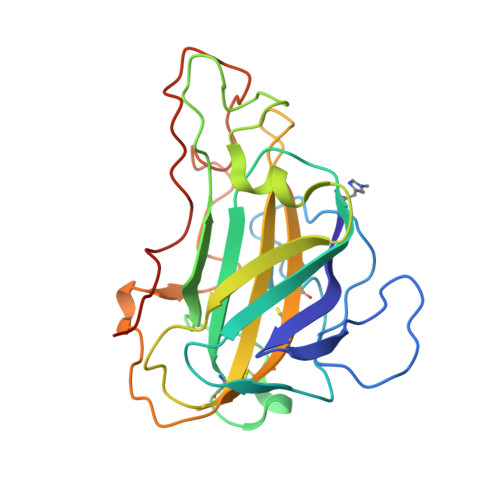
    5UFV

  • PubMed Abstract: 

    Polysaccharide monooxygenases (PMOs) are secreted metalloenzymes that catalyze the oxidative degradation of polysaccharides in a copper-, oxygen-, and reductant-dependent manner. Cellulose-active fungal PMOs degrade cellulosic substrates to be utilized as a carbon source for fungal growth. To gain insight into the PMO mechanism, the role of conserved residues in the copper coordination sphere was investigated. Here, we report active-site hydrogen-bonding motifs in the secondary copper coordination sphere of MtPMO3*, a C1-oxidizing PMO from the ascomycete fungus Myceliophthora thermophila. A series of point substitutions that disrupt this conserved network are used to interrogate its function. Activity assays, in conjunction with EPR spectroscopy, demonstrate that residues H161 and Q167 are involved in stabilizing bound oxygen, and H161 appears to play a role in proton transfer. Additionally, Q167 increases the ligand donor strength of Y169 to the copper via a hydrogen-bonding interaction. Altogether, H161 and Q167 are important for oxygen activation, and the results are suggestive of a copper-oxyl active intermediate.


  • Organizational Affiliation

    Biophysics Graduate Group, University of California, Berkeley , Berkeley, California 94720, United States.


Macromolecules
Find similar proteins by:  (by identity cutoff)  |  3D Structure
Entity ID: 1
MoleculeChains Sequence LengthOrganismDetailsImage
Glycoside hydrolase family 61 protein
A, B, C, D, E
A, B, C, D, E, F
238Thermothelomyces thermophilus ATCC 42464Mutation(s): 0 
Gene Names: MYCTH_92668
UniProt
Find proteins for G2QAB5 (Thermothelomyces thermophilus (strain ATCC 42464 / BCRC 31852 / DSM 1799))
Explore G2QAB5 
Go to UniProtKB:  G2QAB5
Entity Groups  
Sequence Clusters30% Identity50% Identity70% Identity90% Identity95% Identity100% Identity
UniProt GroupG2QAB5
Sequence Annotations
Expand
  • Reference Sequence
Experimental Data & Validation

Experimental Data

  • Method: X-RAY DIFFRACTION
  • Resolution: 2.45 Å
  • R-Value Free: 0.208 
  • R-Value Work: 0.162 
  • R-Value Observed: 0.164 
  • Space Group: P 1 21 1
Unit Cell:
Length ( Å )Angle ( ˚ )
a = 70.937α = 90
b = 134.302β = 92.92
c = 79.402γ = 90
Software Package:
Software NamePurpose
HKL-2000data collection
HKL-2000data reduction
SCALEPACKdata scaling
PHASERphasing
REFMACrefinement
PDB_EXTRACTdata extraction

Structure Validation

View Full Validation Report


Entry History & Funding Information

Deposition Data


Funding OrganizationLocationGrant Number
Energy Biosciences InstituteUnited States022711

Revision History  (Full details and data files)

  • Version 1.0: 2017-03-15
    Type: Initial release
  • Version 1.1: 2017-05-03
    Changes: Database references
  • Version 1.2: 2023-10-04
    Changes: Data collection, Database references, Derived calculations, Refinement description