5UFU

Structure of AMPK bound to activator


Experimental Data Snapshot

  • Method: X-RAY DIFFRACTION
  • Resolution: 3.45 Å
  • R-Value Free: 0.238 
  • R-Value Work: 0.193 
  • R-Value Observed: 0.195 

wwPDB Validation   3D Report Full Report


Ligand Structure Quality Assessment 


This is version 1.0 of the entry. See complete history


Literature

Activation of Skeletal Muscle AMPK Promotes Glucose Disposal and Glucose Lowering in Non-human Primates and Mice.

Cokorinos, E.C.Delmore, J.Reyes, A.R.Albuquerque, B.Kjbsted, R.Jrgensen, N.O.Tran, J.L.Jatkar, A.Cialdea, K.Esquejo, R.M.Meissen, J.Calabrese, M.F.Cordes, J.Moccia, R.Tess, D.Salatto, C.T.Coskran, T.M.Opsahl, A.C.Flynn, D.Blatnik, M.Li, W.Kindt, E.Foretz, M.Viollet, B.Ward, J.Kurumbail, R.G.Kalgutkar, A.S.Wojtaszewski, J.F.P.Cameron, K.O.Miller, R.A.

(2017) Cell Metab 25: 1147-1159.e10

  • DOI: https://doi.org/10.1016/j.cmet.2017.04.010
  • Primary Citation of Related Structures:  
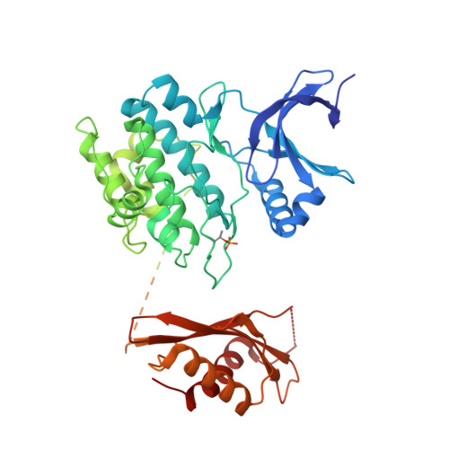
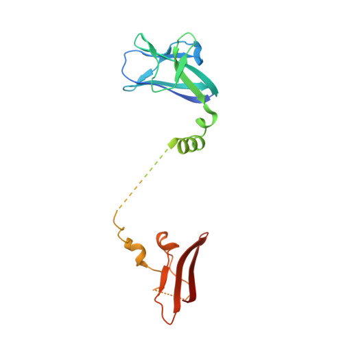
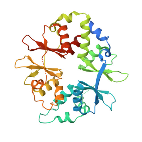
    5UFU

  • PubMed Abstract: 

    The AMP-activated protein kinase (AMPK) is a potential therapeutic target for metabolic diseases based on its reported actions in the liver and skeletal muscle. We evaluated two distinct direct activators of AMPK: a non-selective activator of all AMPK complexes, PF-739, and an activator selective for AMPK β1-containing complexes, PF-249. In cells and animals, both compounds were effective at activating AMPK in hepatocytes, but only PF-739 was capable of activating AMPK in skeletal muscle. In diabetic mice, PF-739, but not PF-249, caused a rapid lowering of plasma glucose levels that was diminished in the absence of skeletal muscle, but not liver, AMPK heterotrimers and was the result of an increase in systemic glucose disposal with no impact on hepatic glucose production. Studies of PF-739 in cynomolgus monkeys confirmed translation of the glucose lowering and established activation of AMPK in skeletal muscle as a potential therapeutic approach to treat diabetic patients.


  • Organizational Affiliation

    Cardiovascular, Metabolic, and Endocrine Diseases Research Unit, Pfizer Inc., Cambridge, MA 02139, USA.


Macromolecules
Find similar proteins by:  (by identity cutoff)  |  3D Structure
Entity ID: 1
MoleculeChains Sequence LengthOrganismDetailsImage
5'-AMP-activated protein kinase catalytic subunit alpha-1503Rattus norvegicusMus musculus
This entity is chimeric
Mutation(s): 0 
Gene Names: Prkaa1Ampk1
EC: 2.7.11.1
UniProt
Find proteins for P54645 (Rattus norvegicus)
Explore P54645 
Go to UniProtKB:  P54645
Entity Groups  
Sequence Clusters30% Identity50% Identity70% Identity90% Identity95% Identity100% Identity
UniProt GroupP54645
Sequence Annotations
Expand
  • Reference Sequence
Find similar proteins by:  (by identity cutoff)  |  3D Structure
Entity ID: 2
MoleculeChains Sequence LengthOrganismDetailsImage
5'-AMP-activated protein kinase subunit beta-1204Rattus norvegicusMutation(s): 1 
Gene Names: Prkab1
UniProt
Find proteins for P80386 (Rattus norvegicus)
Explore P80386 
Go to UniProtKB:  P80386
Entity Groups  
Sequence Clusters30% Identity50% Identity70% Identity90% Identity95% Identity100% Identity
UniProt GroupP80386
Sequence Annotations
Expand
  • Reference Sequence
Find similar proteins by:  (by identity cutoff)  |  3D Structure
Entity ID: 3
MoleculeChains Sequence LengthOrganismDetailsImage
5'-AMP-activated protein kinase subunit gamma-1330Rattus norvegicusMutation(s): 0 
Gene Names: Prkag1
UniProt
Find proteins for P80385 (Rattus norvegicus)
Explore P80385 
Go to UniProtKB:  P80385
Entity Groups  
Sequence Clusters30% Identity50% Identity70% Identity90% Identity95% Identity100% Identity
UniProt GroupP80385
Sequence Annotations
Expand
  • Reference Sequence
Small Molecules
Ligands 6 Unique
IDChains Name / Formula / InChI Key2D Diagram3D Interactions
STU
Query on STU

Download Ideal Coordinates CCD File 
D [auth A]STAUROSPORINE
C28 H26 N4 O3
HKSZLNNOFSGOKW-FYTWVXJKSA-N
85V
Query on 85V

Download Ideal Coordinates CCD File 
E [auth A]1,4:3,6-dianhydro-2-O-(6-chloro-5-{4-[1-(hydroxymethyl)cyclopropyl]phenyl}-1H-benzimidazol-2-yl)-D-mannitol
C23 H23 Cl N2 O5
IMVYDTHPRTUISR-XRXFAXGQSA-N
ADP
Query on ADP

Download Ideal Coordinates CCD File 
L [auth C]ADENOSINE-5'-DIPHOSPHATE
C10 H15 N5 O10 P2
XTWYTFMLZFPYCI-KQYNXXCUSA-N
AMP
Query on AMP

Download Ideal Coordinates CCD File 
J [auth C],
K [auth C]
ADENOSINE MONOPHOSPHATE
C10 H14 N5 O7 P
UDMBCSSLTHHNCD-KQYNXXCUSA-N
SO4
Query on SO4

Download Ideal Coordinates CCD File 
I [auth A],
M [auth C]
SULFATE ION
O4 S
QAOWNCQODCNURD-UHFFFAOYSA-L
CL
Query on CL

Download Ideal Coordinates CCD File 
F [auth A],
G [auth A],
H [auth A]
CHLORIDE ION
Cl
VEXZGXHMUGYJMC-UHFFFAOYSA-M
Modified Residues  1 Unique
IDChains TypeFormula2D DiagramParent
TPO
Query on TPO
A
L-PEPTIDE LINKINGC4 H10 N O6 PTHR
Experimental Data & Validation

Experimental Data

  • Method: X-RAY DIFFRACTION
  • Resolution: 3.45 Å
  • R-Value Free: 0.238 
  • R-Value Work: 0.193 
  • R-Value Observed: 0.195 
  • Space Group: P 61 2 2
Unit Cell:
Length ( Å )Angle ( ˚ )
a = 124.264α = 90
b = 124.264β = 90
c = 401.569γ = 120
Software Package:
Software NamePurpose
XSCALEdata scaling
PHASERphasing
BUSTERrefinement
PDB_EXTRACTdata extraction
XDSdata reduction
XDSdata scaling
PHASERphasing

Structure Validation

View Full Validation Report


Ligand Structure Quality Assessment 


Entry History 

Deposition Data

Revision History  (Full details and data files)

  • Version 1.0: 2017-05-17
    Type: Initial release