5UFM

Crystal structure of Burkholderia thailandensis 1,6-didemethyltoxoflavin-N1-methyltransferase with bound 1,6-didemethyltoxoflavin and S-adenosylhomocysteine


Experimental Data Snapshot

  • Method: X-RAY DIFFRACTION
  • Resolution: 1.77 Å
  • R-Value Free: 0.184 
  • R-Value Work: 0.152 
  • R-Value Observed: 0.154 

wwPDB Validation   3D Report Full Report


Ligand Structure Quality Assessment 


This is version 1.3 of the entry. See complete history


Literature

Biochemical Characterization and Structural Basis of Reactivity and Regioselectivity Differences between Burkholderia thailandensis and Burkholderia glumae 1,6-Didesmethyltoxoflavin N-Methyltransferase.

Fenwick, M.K.Almabruk, K.H.Ealick, S.E.Begley, T.P.Philmus, B.

(2017) Biochemistry 56: 3934-3944

  • DOI: https://doi.org/10.1021/acs.biochem.7b00476
  • Primary Citation of Related Structures:  
    5UFM, 5UFN

  • PubMed Abstract: 

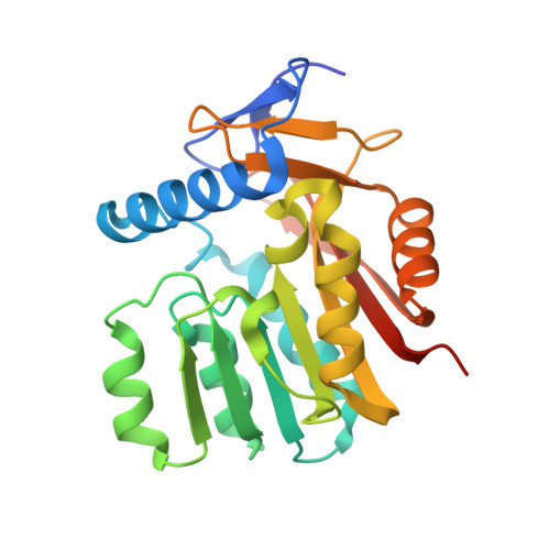
    Burkholderia glumae converts the guanine base of guanosine triphosphate into an azapteridine and methylates both the pyrimidine and triazine rings to make toxoflavin. Strains of Burkholderia thailandensis and Burkholderia pseudomallei have a gene cluster encoding seven putative biosynthetic enzymes that resembles the toxoflavin gene cluster. Four of the enzymes are similar in sequence to BgToxBCDE, which have been proposed to make 1,6-didesmethyltoxoflavin (1,6-DDMT). One of the remaining enzymes, BthII1283 in B. thailandensis E264, is a predicted S-adenosylmethionine (SAM)-dependent N-methyltransferase that shows a low level of sequence identity to BgToxA, which sequentially methylates N6 and N1 of 1,6-DDMT to form toxoflavin. Here we show that, unlike BgToxA, BthII1283 catalyzes a single methyl transfer to N1 of 1,6-DDMT in vitro. In addition, we investigated the differences in reactivity and regioselectivity by determining crystal structures of BthII1283 with bound S-adenosylhomocysteine (SAH) or 1,6-DDMT and SAH. BthII1283 contains a class I methyltransferase fold and three unique extensions used for 1,6-DDMT recognition. The active site structure suggests that 1,6-DDMT is bound in a reduced form. The plane of the azapteridine ring system is orthogonal to its orientation in BgToxA. In BthII1283, the modeled SAM methyl group is directed toward the p orbital of N1, whereas in BgToxA, it is first directed toward an sp 2 orbital of N6 and then toward an sp 2 orbital of N1 after planar rotation of the azapteridine ring system. Furthermore, in BthII1283, N1 is hydrogen bonded to a histidine residue whereas BgToxA does not supply an obvious basic residue for either N6 or N1 methylation.


  • Organizational Affiliation

    Department of Chemistry and Chemical Biology, Cornell University , Ithaca, New York 14853, United States.


Macromolecules
Find similar proteins by:  (by identity cutoff)  |  3D Structure
Entity ID: 1
MoleculeChains Sequence LengthOrganismDetailsImage
Methyltransferase domain protein
A, B
238Burkholderia thailandensis E264Mutation(s): 0 
Gene Names: BTH_II1283DR63_4497
UniProt
Find proteins for Q2T5S0 (Burkholderia thailandensis (strain ATCC 700388 / DSM 13276 / CCUG 48851 / CIP 106301 / E264))
Explore Q2T5S0 
Go to UniProtKB:  Q2T5S0
Entity Groups  
Sequence Clusters30% Identity50% Identity70% Identity90% Identity95% Identity100% Identity
UniProt GroupQ2T5S0
Sequence Annotations
Expand
  • Reference Sequence
Small Molecules
Ligands 4 Unique
IDChains Name / Formula / InChI Key2D Diagram3D Interactions
SAH
Query on SAH

Download Ideal Coordinates CCD File 
C [auth A],
N [auth B]
S-ADENOSYL-L-HOMOCYSTEINE
C14 H20 N6 O5 S
ZJUKTBDSGOFHSH-WFMPWKQPSA-N
EPE
Query on EPE

Download Ideal Coordinates CCD File 
J [auth A],
K [auth A]
4-(2-HYDROXYETHYL)-1-PIPERAZINE ETHANESULFONIC ACID
C8 H18 N2 O4 S
JKMHFZQWWAIEOD-UHFFFAOYSA-N
AZ8
Query on AZ8

Download Ideal Coordinates CCD File 
D [auth A],
O [auth B]
pyrimido[5,4-e][1,2,4]triazine-5,7(6H,8H)-dione
C5 H3 N5 O2
IDJLTUNWTSUIHO-UHFFFAOYSA-N
SO4
Query on SO4

Download Ideal Coordinates CCD File 
E [auth A]
F [auth A]
G [auth A]
H [auth A]
I [auth A]
E [auth A],
F [auth A],
G [auth A],
H [auth A],
I [auth A],
L [auth A],
M [auth B],
P [auth B],
Q [auth B],
R [auth B],
S [auth B]
SULFATE ION
O4 S
QAOWNCQODCNURD-UHFFFAOYSA-L
Experimental Data & Validation

Experimental Data

  • Method: X-RAY DIFFRACTION
  • Resolution: 1.77 Å
  • R-Value Free: 0.184 
  • R-Value Work: 0.152 
  • R-Value Observed: 0.154 
  • Space Group: P 61
Unit Cell:
Length ( Å )Angle ( ˚ )
a = 112.698α = 90
b = 112.698β = 90
c = 78.367γ = 120
Software Package:
Software NamePurpose
HKL-2000data processing
PHENIXrefinement
Cootmodel building

Structure Validation

View Full Validation Report


Ligand Structure Quality Assessment 


Entry History & Funding Information

Deposition Data


Funding OrganizationLocationGrant Number
National Institutes of Health/National Institute of General Medical Sciences (NIH/NIGMS)United StatesGM103403
National Institutes of Health/National Institute of General Medical Sciences (NIH/NIGMS)United StatesGM103485
Department of Energy (DOE, United States)United StatesDE-AC02-06CH11357
Oregon State University College of PharmacyUnited States--

Revision History  (Full details and data files)

  • Version 1.0: 2017-12-13
    Type: Initial release
  • Version 1.1: 2018-12-26
    Changes: Data collection, Database references
  • Version 1.2: 2019-12-04
    Changes: Author supporting evidence
  • Version 1.3: 2024-03-06
    Changes: Data collection, Database references