5UFL

Crystal structure of a CIP2A core domain


Experimental Data Snapshot

  • Method: X-RAY DIFFRACTION
  • Resolution: 3.00 Å
  • R-Value Free: 0.281 
  • R-Value Work: 0.256 
  • R-Value Observed: 0.257 

wwPDB Validation   3D Report Full Report


This is version 1.4 of the entry. See complete history


Literature

Oncoprotein CIP2A is stabilized via interaction with tumor suppressor PP2A/B56.

Wang, J.Okkeri, J.Pavic, K.Wang, Z.Kauko, O.Halonen, T.Sarek, G.Ojala, P.M.Rao, Z.Xu, W.Westermarck, J.

(2017) EMBO Rep 18: 437-450

  • DOI: https://doi.org/10.15252/embr.201642788
  • Primary Citation of Related Structures:  
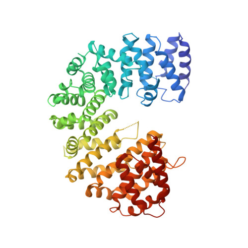
    5UFL

  • PubMed Abstract: 

    Protein phosphatase 2A (PP2A) is a critical human tumor suppressor. Cancerous inhibitor of PP2A (CIP2A) supports the activity of several critical cancer drivers (Akt, MYC, E2F1) and promotes malignancy in most cancer types via PP2A inhibition. However, the 3D structure of CIP2A has not been solved, and it remains enigmatic how it interacts with PP2A. Here, we show by yeast two-hybrid assays, and subsequent validation experiments, that CIP2A forms homodimers. The homodimerization of CIP2A is confirmed by solving the crystal structure of an N-terminal CIP2A fragment (amino acids 1-560) at 3.0 Å resolution, and by subsequent structure-based mutational analyses of the dimerization interface. We further describe that the CIP2A dimer interacts with the PP2A subunits B56α and B56γ. CIP2A binds to the B56 proteins via a conserved N-terminal region, and dimerization promotes B56 binding. Intriguingly, inhibition of either CIP2A dimerization or B56α/γ expression destabilizes CIP2A, indicating opportunities for controlled degradation. These results provide the first structure-function analysis of the interaction of CIP2A with PP2A/B56 and have direct implications for its targeting in cancer therapy.


  • Organizational Affiliation

    Department of Biological Structure, University of Washington, Seattle, WA, USA.


Macromolecules
Find similar proteins by:  (by identity cutoff)  |  3D Structure
Entity ID: 1
MoleculeChains Sequence LengthOrganismDetailsImage
Protein CIP2A
A, B
561Homo sapiensMutation(s): 0 
Gene Names: KIAA1524CIP2A
UniProt & NIH Common Fund Data Resources
Find proteins for Q8TCG1 (Homo sapiens)
Explore Q8TCG1 
Go to UniProtKB:  Q8TCG1
PHAROS:  Q8TCG1
GTEx:  ENSG00000163507 
Entity Groups  
Sequence Clusters30% Identity50% Identity70% Identity90% Identity95% Identity100% Identity
UniProt GroupQ8TCG1
Sequence Annotations
Expand
  • Reference Sequence
Small Molecules
Ligands 1 Unique
IDChains Name / Formula / InChI Key2D Diagram3D Interactions
ZN
Query on ZN

Download Ideal Coordinates CCD File 
C [auth A]ZINC ION
Zn
PTFCDOFLOPIGGS-UHFFFAOYSA-N
Experimental Data & Validation

Experimental Data

  • Method: X-RAY DIFFRACTION
  • Resolution: 3.00 Å
  • R-Value Free: 0.281 
  • R-Value Work: 0.256 
  • R-Value Observed: 0.257 
  • Space Group: P 65
Unit Cell:
Length ( Å )Angle ( ˚ )
a = 153.276α = 90
b = 153.276β = 90
c = 105.44γ = 120
Software Package:
Software NamePurpose
HKL-2000data scaling
REFMACrefinement
PDB_EXTRACTdata extraction
HKL-2000data reduction
PHASERphasing
DENZOdata reduction
SCALEPACKdata scaling

Structure Validation

View Full Validation Report


Entry History 

Deposition Data

Revision History  (Full details and data files)

  • Version 1.0: 2017-02-15
    Type: Initial release
  • Version 1.1: 2017-02-22
    Changes: Database references
  • Version 1.2: 2017-03-15
    Changes: Database references
  • Version 1.3: 2017-11-22
    Changes: Refinement description
  • Version 1.4: 2024-03-06
    Changes: Data collection, Database references, Derived calculations, Refinement description