5UC1

Structural Analysis of Glucocorticoid Receptor beta Ligand Binding Domain Complexed with Glucocorticoid Antagonist RU-486: Implication of Helix 12 in Antagonism


Experimental Data Snapshot

  • Method: X-RAY DIFFRACTION
  • Resolution: 2.35 Å
  • R-Value Free: 0.254 
  • R-Value Work: 0.204 
  • R-Value Observed: 0.206 

wwPDB Validation   3D Report Full Report


Ligand Structure Quality Assessment 


This is version 1.2 of the entry. See complete history


Literature

Probing Dominant Negative Behavior of Glucocorticoid Receptor beta through a Hybrid Structural and Biochemical Approach.

Min, J.Perera, L.Krahn, J.M.Jewell, C.M.Moon, A.F.Cidlowski, J.A.Pedersen, L.C.

(2018) Mol Cell Biol 

  • DOI: https://doi.org/10.1128/MCB.00453-17
  • Primary Citation of Related Structures:  
    5UC1

  • PubMed Abstract: 

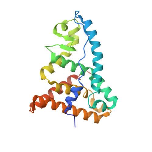
    Glucocorticoid receptor β (GRβ) is associated with glucocorticoid resistance via dominant negative regulation of GRα. To better understand how GRβ functions as a dominant negative inhibitor of GRα at a molecular level, we determined the crystal structure of the ligand binding domain of GRβ complexed with the antagonist RU-486. The structure reveals that GRβ binds RU-486 in the same ligand binding pocket as GRα, and the unique C-terminal amino acids of GRβ are mostly disordered. Binding energy analysis suggests that these C-terminal residues of GRβ do not contribute to RU-486 binding. Intriguingly, the GRβ/RU-486 complex binds corepressor peptide with affinity similar to that of a GRα/RU-486 complex, despite the lack of helix 12. Our biophysical and biochemical analyses reveal that in the presence of RU-486, GRβ is found in a conformation that favors corepressor binding, potentially antagonizing GRα function. This study thus presents an unexpected molecular mechanism by which GRβ could repress transcription.


  • Organizational Affiliation

    Genome Integrity and Structural Biology Laboratory, National Institute of Environmental Health Sciences, National Institutes of Health, Research Triangle Park, North Carolina, USA.


Macromolecules
Find similar proteins by:  (by identity cutoff)  |  3D Structure
Entity ID: 1
MoleculeChains Sequence LengthOrganismDetailsImage
Glucocorticoid receptor
A, B
222Heterocephalus glaberMutation(s): 0 
Gene Names: GW7_10599
UniProt
Find proteins for G5AQS2 (Heterocephalus glaber)
Explore G5AQS2 
Go to UniProtKB:  G5AQS2
Entity Groups  
Sequence Clusters30% Identity50% Identity70% Identity90% Identity95% Identity100% Identity
UniProt GroupG5AQS2
Sequence Annotations
Expand
  • Reference Sequence
Small Molecules
Ligands 5 Unique
IDChains Name / Formula / InChI Key2D Diagram3D Interactions
CPS
Query on CPS

Download Ideal Coordinates CCD File 
D [auth A],
E [auth A],
L [auth B],
M [auth B]
3-[(3-CHOLAMIDOPROPYL)DIMETHYLAMMONIO]-1-PROPANESULFONATE
C32 H58 N2 O7 S
UMCMPZBLKLEWAF-BCTGSCMUSA-N
486
Query on 486

Download Ideal Coordinates CCD File 
C [auth A],
K [auth B]
11-(4-DIMETHYLAMINO-PHENYL)-17-HYDROXY-13-METHYL-17-PROP-1-YNYL-1,2,6,7,8,11,12,13,14,15,16,17-DODEC AHYDRO-CYCLOPENTA[A]PHENANTHREN-3-ONE
C29 H35 N O2
VKHAHZOOUSRJNA-GCNJZUOMSA-N
MPD
Query on MPD

Download Ideal Coordinates CCD File 
H [auth A],
P [auth B]
(4S)-2-METHYL-2,4-PENTANEDIOL
C6 H14 O2
SVTBMSDMJJWYQN-YFKPBYRVSA-N
EDO
Query on EDO

Download Ideal Coordinates CCD File 
I [auth A],
J [auth A],
Q [auth B],
R [auth B],
S [auth B]
1,2-ETHANEDIOL
C2 H6 O2
LYCAIKOWRPUZTN-UHFFFAOYSA-N
CL
Query on CL

Download Ideal Coordinates CCD File 
F [auth A],
G [auth A],
N [auth B],
O [auth B]
CHLORIDE ION
Cl
VEXZGXHMUGYJMC-UHFFFAOYSA-M
Binding Affinity Annotations 
IDSourceBinding Affinity
486 BindingDB:  5UC1 Ki: min: 0.09, max: 169 (nM) from 18 assay(s)
Kd: 15 (nM) from 1 assay(s)
IC50: min: 8.00e-3, max: 33 (nM) from 33 assay(s)
EC50: min: 0.6, max: 2500 (nM) from 6 assay(s)
Experimental Data & Validation

Experimental Data

  • Method: X-RAY DIFFRACTION
  • Resolution: 2.35 Å
  • R-Value Free: 0.254 
  • R-Value Work: 0.204 
  • R-Value Observed: 0.206 
  • Space Group: P 61
Unit Cell:
Length ( Å )Angle ( ˚ )
a = 48.361α = 90
b = 48.361β = 90
c = 385.802γ = 120
Software Package:
Software NamePurpose
HKL-2000data collection
PHASERphasing
PHENIXrefinement
PDB_EXTRACTdata extraction
HKL-2000data reduction
HKL-2000data scaling
HKLdata scaling

Structure Validation

View Full Validation Report


Ligand Structure Quality Assessment 


Entry History 

Deposition Data

Revision History  (Full details and data files)

  • Version 1.0: 2017-12-27
    Type: Initial release
  • Version 1.1: 2018-06-20
    Changes: Data collection, Database references
  • Version 1.2: 2023-10-04
    Changes: Data collection, Database references, Refinement description, Structure summary