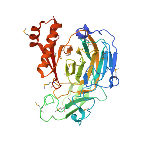
2.75 Angstrom Resolution Crystal Structure of Acetamidase from Yersinia enterocolitica.
Minasov, G., Shuvalova, L., Flores, K., Dubrovska, I., Grimshaw, S., Kwon, K., Anderson, W.F., Center for Structural Genomics of Infectious Diseases (CSGID)To be published.