5U36

Crystal Structure Of A Mutant M. Jannashii Tyrosyl-tRNA Synthetase


Experimental Data Snapshot

  • Method: X-RAY DIFFRACTION
  • Resolution: 3.03 Å
  • R-Value Free: 0.282 
  • R-Value Work: 0.201 
  • R-Value Observed: 0.205 

wwPDB Validation   3D Report Full Report


This is version 1.5 of the entry. See complete history


Literature

Genetically encoding phosphotyrosine and its nonhydrolyzable analog in bacteria.

Luo, X.Fu, G.Wang, R.E.Zhu, X.Zambaldo, C.Liu, R.Liu, T.Lyu, X.Du, J.Xuan, W.Yao, A.Reed, S.A.Kang, M.Zhang, Y.Guo, H.Huang, C.Yang, P.Y.Wilson, I.A.Schultz, P.G.Wang, F.

(2017) Nat Chem Biol 13: 845-849

  • DOI: https://doi.org/10.1038/nchembio.2405
  • Primary Citation of Related Structures:  
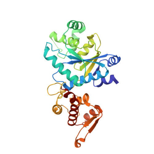
    5U36

  • PubMed Abstract: 

    Tyrosine phosphorylation is a common protein post-translational modification that plays a critical role in signal transduction and the regulation of many cellular processes. Using a propeptide strategy to increase cellular uptake of O-phosphotyrosine (pTyr) and its nonhydrolyzable analog 4-phosphomethyl-L-phenylalanine (Pmp), we identified an orthogonal aminoacyl-tRNA synthetase-tRNA pair that allows site-specific incorporation of both pTyr and Pmp into recombinant proteins in response to the amber stop codon in Escherichia coli in good yields. The X-ray structure of the synthetase reveals a reconfigured substrate-binding site, formed by nonconservative mutations and substantial local structural perturbations. We demonstrate the utility of this method by introducing Pmp into a putative phosphorylation site and determining the affinities of the individual variants for the substrate 3BP2. In summary, this work provides a useful recombinant tool to dissect the biological functions of tyrosine phosphorylation at specific sites in the proteome.


  • Organizational Affiliation

    Department of Chemistry, The Scripps Research Institute, La Jolla, California, USA.


Macromolecules
Find similar proteins by:  (by identity cutoff)  |  3D Structure
Entity ID: 1
MoleculeChains Sequence LengthOrganismDetailsImage
Tyrosine--tRNA ligase
A, B
314Methanocaldococcus jannaschii DSM 2661Mutation(s): 6 
Gene Names: tyrSMJ0389
EC: 6.1.1.1
UniProt
Find proteins for Q57834 (Methanocaldococcus jannaschii (strain ATCC 43067 / DSM 2661 / JAL-1 / JCM 10045 / NBRC 100440))
Explore Q57834 
Go to UniProtKB:  Q57834
Entity Groups  
Sequence Clusters30% Identity50% Identity70% Identity90% Identity95% Identity100% Identity
UniProt GroupQ57834
Sequence Annotations
Expand
  • Reference Sequence
Experimental Data & Validation

Experimental Data

  • Method: X-RAY DIFFRACTION
  • Resolution: 3.03 Å
  • R-Value Free: 0.282 
  • R-Value Work: 0.201 
  • R-Value Observed: 0.205 
  • Space Group: P 1 21 1
Unit Cell:
Length ( Å )Angle ( ˚ )
a = 58.634α = 90
b = 130.559β = 117.71
c = 59.956γ = 90
Software Package:
Software NamePurpose
REFMACrefinement
DENZOdata reduction
SCALEPACKdata scaling
PHASERphasing

Structure Validation

View Full Validation Report


Entry History & Funding Information

Deposition Data


Funding OrganizationLocationGrant Number
National Institutes of Health/National Institute of General Medical Sciences (NIH/NIGMS)United StatesGM62159-14

Revision History  (Full details and data files)

  • Version 1.0: 2017-06-07
    Type: Initial release
  • Version 1.1: 2017-06-28
    Changes: Database references
  • Version 1.2: 2017-09-06
    Changes: Database references
  • Version 1.3: 2017-09-20
    Changes: Author supporting evidence
  • Version 1.4: 2020-01-01
    Changes: Author supporting evidence
  • Version 1.5: 2023-10-04
    Changes: Data collection, Database references, Refinement description