5U2H

Crystal structure of the ATP-gated P2X7 ion channel bound to ATP and allosteric antagonist A804598

  • Classification: MEMBRANE PROTEIN
  • Organism(s): Ailuropoda melanoleuca
  • Expression System: Spodoptera frugiperda
  • Mutation(s): Yes 
  • Membrane Protein: Yes  OPMPDBTM

  • Deposited: 2016-11-30 Released: 2017-01-04 
  • Deposition Author(s): Karasawa, A., Kawate, T.
  • Funding Organization(s): National Institutes of Health/National Institute of General Medical Sciences (NIH/NIGMS), National Institutes of Health/National Institute of Neurological Disorders and Stroke (NIH/NINDS)

Experimental Data Snapshot

  • Method: X-RAY DIFFRACTION
  • Resolution: 3.90 Å
  • R-Value Free: 0.386 
  • R-Value Work: 0.334 
  • R-Value Observed: 0.337 

wwPDB Validation   3D Report Full Report


Ligand Structure Quality Assessment 


This is version 2.1 of the entry. See complete history


Literature

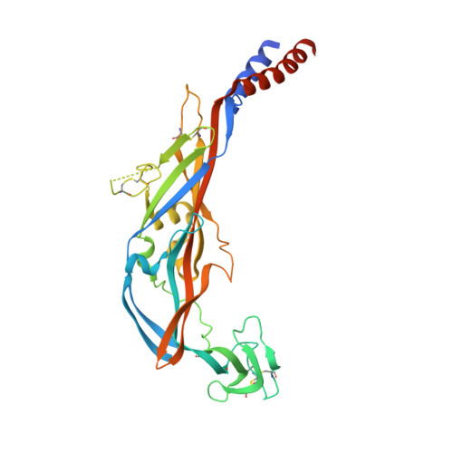
Structural basis for subtype-specific inhibition of the P2X7 receptor.

Karasawa, A.Kawate, T.

(2016) Elife 5

  • DOI: https://doi.org/10.7554/eLife.22153
  • Primary Citation of Related Structures:  
    5U1L, 5U1U, 5U1V, 5U1W, 5U1X, 5U1Y, 5U2H

  • PubMed Abstract: 

    The P2X7 receptor is a non-selective cation channel activated by extracellular adenosine triphosphate (ATP). Chronic activation of P2X7 underlies many health problems such as pathologic pain, yet we lack effective antagonists due to poorly understood mechanisms of inhibition. Here we present crystal structures of a mammalian P2X7 receptor complexed with five structurally-unrelated antagonists. Unexpectedly, these drugs all bind to an allosteric site distinct from the ATP-binding pocket in a groove formed between two neighboring subunits. This novel drug-binding pocket accommodates a diversity of small molecules mainly through hydrophobic interactions. Functional assays propose that these compounds allosterically prevent narrowing of the drug-binding pocket and the turret-like architecture during channel opening, which is consistent with a site of action distal to the ATP-binding pocket. These novel mechanistic insights will facilitate the development of P2X7-specific drugs for treating human diseases.


  • Organizational Affiliation

    Department of Molecular Medicine, Cornell University, Ithaca, United States.


Macromolecules
Find similar proteins by:  (by identity cutoff)  |  3D Structure
Entity ID: 1
MoleculeChains Sequence LengthOrganismDetailsImage
P2X purinoceptor
A, B
342Ailuropoda melanoleucaMutation(s): 5 
Gene Names: P2RX7
Membrane Entity: Yes 
UniProt
Find proteins for G1M6C4 (Ailuropoda melanoleuca)
Explore G1M6C4 
Go to UniProtKB:  G1M6C4
Entity Groups  
Sequence Clusters30% Identity50% Identity70% Identity90% Identity95% Identity100% Identity
UniProt GroupG1M6C4
Sequence Annotations
Expand
  • Reference Sequence
Oligosaccharides

Help

Entity ID: 2
MoleculeChains Length2D Diagram Glycosylation3D Interactions
2-acetamido-2-deoxy-beta-D-glucopyranose-(1-4)-2-acetamido-2-deoxy-beta-D-glucopyranose
C, D
2N-Glycosylation
Glycosylation Resources
GlyTouCan:  G42666HT
GlyCosmos:  G42666HT
GlyGen:  G42666HT
Small Molecules
Binding Affinity Annotations 
IDSourceBinding Affinity
ATP BindingDB:  5U2H EC50: 7.80e+5 (nM) from 1 assay(s)
Experimental Data & Validation

Experimental Data

  • Method: X-RAY DIFFRACTION
  • Resolution: 3.90 Å
  • R-Value Free: 0.386 
  • R-Value Work: 0.334 
  • R-Value Observed: 0.337 
  • Space Group: P 21 3
Unit Cell:
Length ( Å )Angle ( ˚ )
a = 167.618α = 90
b = 167.618β = 90
c = 167.618γ = 90
Software Package:
Software NamePurpose
PHENIXrefinement
Aimlessdata scaling
PHASERphasing
PDB_EXTRACTdata extraction
XSCALEdata scaling

Structure Validation

View Full Validation Report


Ligand Structure Quality Assessment 


Entry History & Funding Information

Deposition Data


Funding OrganizationLocationGrant Number
National Institutes of Health/National Institute of General Medical Sciences (NIH/NIGMS)United StatesGM114379
National Institutes of Health/National Institute of Neurological Disorders and Stroke (NIH/NINDS)United StatesNS072869

Revision History  (Full details and data files)

  • Version 1.0: 2017-01-04
    Type: Initial release
  • Version 1.1: 2017-09-20
    Changes: Author supporting evidence, Refinement description
  • Version 1.2: 2019-12-18
    Changes: Author supporting evidence
  • Version 2.0: 2020-07-29
    Type: Remediation
    Reason: Carbohydrate remediation
    Changes: Atomic model, Data collection, Derived calculations, Structure summary
  • Version 2.1: 2024-04-03
    Changes: Data collection, Database references, Refinement description, Structure summary