5U0L

X-ray crystal structure of fatty aldehyde dehydrogenase enzymes from Marinobacter aquaeolei VT8 complexed with a substrate


Experimental Data Snapshot

  • Method: X-RAY DIFFRACTION
  • Resolution: 2.29 Å
  • R-Value Free: 0.270 
  • R-Value Work: 0.214 
  • R-Value Observed: 0.217 

wwPDB Validation   3D Report Full Report


Ligand Structure Quality Assessment 


This is version 1.4 of the entry. See complete history


Literature

Five Fatty Aldehyde Dehydrogenase Enzymes from Marinobacter and Acinetobacter spp. and Structural Insights into the Aldehyde Binding Pocket.

Bertram, J.H.Mulliner, K.M.Shi, K.Plunkett, M.H.Nixon, P.Serratore, N.A.Douglas, C.J.Aihara, H.Barney, B.M.

(2017) Appl Environ Microbiol 83

  • DOI: https://doi.org/10.1128/AEM.00018-17
  • Primary Citation of Related Structures:  
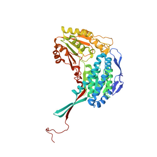
    5U0L, 5U0M

  • PubMed Abstract: 

    Enzymes involved in lipid biosynthesis and metabolism play an important role in energy conversion and storage and in the function of structural components such as cell membranes. The fatty aldehyde dehydrogenase (FAldDH) plays a central function in the metabolism of lipid intermediates, oxidizing fatty aldehydes to the corresponding fatty acid and competing with pathways that would further reduce the fatty aldehydes to fatty alcohols or require the fatty aldehydes to produce alkanes. In this report, the genes for four putative FAldDH enzymes from Marinobacter aquaeolei VT8 and an additional enzyme from Acinetobacter baylyi were heterologously expressed in Escherichia coli and shown to display FAldDH activity. Five enzymes (Maqu_0438, Maqu_3316, Maqu_3410, Maqu_3572, and the enzyme reported under RefSeq accession no. WP_004927398) were found to act on aldehydes ranging from acetaldehyde to hexadecanal and also acted on the unsaturated long-chain palmitoleyl and oleyl aldehydes. A comparison of the specificities of these enzymes with various aldehydes is presented. Crystallization trials yielded diffraction-quality crystals of one particular FAldDH (Maqu_3316) from M. aquaeolei VT8. Crystals were independently treated with both the NAD + cofactor and the aldehyde substrate decanal, revealing specific details of the likely substrate binding pocket for this class of enzymes. A likely model for how catalysis by the enzyme is accomplished is also provided. IMPORTANCE This study provides a comparison of multiple enzymes with the ability to oxidize fatty aldehydes to fatty acids and provides a likely picture of how the fatty aldehyde and NAD + are bound to the enzyme to facilitate catalysis. Based on the information obtained from this structural analysis and comparisons of specificities for the five enzymes that were characterized, correlations to the potential roles played by specific residues within the structure may be drawn.


  • Organizational Affiliation

    Department of Bioproducts and Biosystems Engineering, University of Minnesota, St. Paul, Minnesota, USA.


Macromolecules
Find similar proteins by:  (by identity cutoff)  |  3D Structure
Entity ID: 1
MoleculeChains Sequence LengthOrganismDetailsImage
N-succinylglutamate 5-semialdehyde dehydrogenase
A, B
497Marinobacter nauticus VT8Mutation(s): 0 
Gene Names: astDMaqu_3316
EC: 1.2.1.71
UniProt
Find proteins for A1U5W8 (Marinobacter nauticus (strain ATCC 700491 / DSM 11845 / VT8))
Explore A1U5W8 
Go to UniProtKB:  A1U5W8
Entity Groups  
Sequence Clusters30% Identity50% Identity70% Identity90% Identity95% Identity100% Identity
UniProt GroupA1U5W8
Sequence Annotations
Expand
  • Reference Sequence
Small Molecules
Ligands 5 Unique
IDChains Name / Formula / InChI Key2D Diagram3D Interactions
8YP
Query on 8YP

Download Ideal Coordinates CCD File 
I [auth A]decanal
C10 H20 O
KSMVZQYAVGTKIV-UHFFFAOYSA-N
MPD
Query on MPD

Download Ideal Coordinates CCD File 
L [auth B],
M [auth B]
(4S)-2-METHYL-2,4-PENTANEDIOL
C6 H14 O2
SVTBMSDMJJWYQN-YFKPBYRVSA-N
PO4
Query on PO4

Download Ideal Coordinates CCD File 
O [auth B]PHOSPHATE ION
O4 P
NBIIXXVUZAFLBC-UHFFFAOYSA-K
EDO
Query on EDO

Download Ideal Coordinates CCD File 
C [auth A]
D [auth A]
E [auth A]
F [auth A]
G [auth A]
C [auth A],
D [auth A],
E [auth A],
F [auth A],
G [auth A],
H [auth A],
J [auth A],
K [auth A]
1,2-ETHANEDIOL
C2 H6 O2
LYCAIKOWRPUZTN-UHFFFAOYSA-N
CL
Query on CL

Download Ideal Coordinates CCD File 
N [auth B]CHLORIDE ION
Cl
VEXZGXHMUGYJMC-UHFFFAOYSA-M
Experimental Data & Validation

Experimental Data

  • Method: X-RAY DIFFRACTION
  • Resolution: 2.29 Å
  • R-Value Free: 0.270 
  • R-Value Work: 0.214 
  • R-Value Observed: 0.217 
  • Space Group: P 41 21 2
Unit Cell:
Length ( Å )Angle ( ˚ )
a = 98.71α = 90
b = 98.71β = 90
c = 254.725γ = 90
Software Package:
Software NamePurpose
PHENIXrefinement
XDSdata reduction
Aimlessdata scaling
PHASERphasing

Structure Validation

View Full Validation Report


Ligand Structure Quality Assessment 


Entry History & Funding Information

Deposition Data


Funding OrganizationLocationGrant Number
National Institutes of Health/National Institute of General Medical Sciences (NIH/NIGMS)United StatesGM118047
National Science Foundation (NSF, United States)United StatesCBET-1437758
National Science Foundation (NSF, United States)United States0968781

Revision History  (Full details and data files)

  • Version 1.0: 2017-04-26
    Type: Initial release
  • Version 1.1: 2017-06-14
    Changes: Database references
  • Version 1.2: 2017-09-13
    Changes: Author supporting evidence
  • Version 1.3: 2019-11-27
    Changes: Author supporting evidence
  • Version 1.4: 2023-10-04
    Changes: Data collection, Database references, Refinement description