5TWX

Crystal Structure of BRD9 bromodomain


Experimental Data Snapshot

  • Method: X-RAY DIFFRACTION
  • Resolution: 2.55 Å
  • R-Value Free: 0.280 
  • R-Value Work: 0.233 
  • R-Value Observed: 0.235 

wwPDB Validation   3D Report Full Report


Ligand Structure Quality Assessment 


This is version 1.1 of the entry. See complete history


Literature

Degradation of the BAF Complex Factor BRD9 by Heterobifunctional Ligands.

Remillard, D.Buckley, D.L.Paulk, J.Brien, G.L.Sonnett, M.Seo, H.S.Dastjerdi, S.Wuhr, M.Dhe-Paganon, S.Armstrong, S.A.Bradner, J.E.

(2017) Angew Chem Int Ed Engl 56: 5738-5743

  • DOI: https://doi.org/10.1002/anie.201611281
  • Primary Citation of Related Structures:  
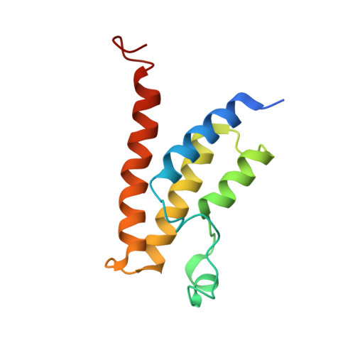
    5TWX

  • PubMed Abstract: 

    The bromodomain-containing protein BRD9, a subunit of the human BAF (SWI/SNF) nucleosome remodeling complex, has emerged as an attractive therapeutic target in cancer. Despite the development of chemical probes targeting the BRD9 bromodomain, there is a limited understanding of BRD9 function beyond acetyl-lysine recognition. We have therefore created the first BRD9-directed chemical degraders, through iterative design and testing of heterobifunctional ligands that bridge the BRD9 bromodomain and the cereblon E3 ubiquitin ligase complex. Degraders of BRD9 exhibit markedly enhanced potency compared to parental ligands (10- to 100-fold). Parallel study of degraders with divergent BRD9-binding chemotypes in models of acute myeloid leukemia resolves bromodomain polypharmacology in this emerging drug class. Together, these findings reveal the tractability of non-BET bromodomain containing proteins to chemical degradation, and highlight lead compound dBRD9 as a tool for the study of BRD9.


  • Organizational Affiliation

    Department of Medical Oncology, Dana-Farber Cancer Institute, Boston, MA, USA.


Macromolecules
Find similar proteins by:  (by identity cutoff)  |  3D Structure
Entity ID: 1
MoleculeChains Sequence LengthOrganismDetailsImage
Bromodomain-containing protein 9
A, B, C, D
121Homo sapiensMutation(s): 0 
Gene Names: BRD9UNQ3040/PRO9856
UniProt & NIH Common Fund Data Resources
Find proteins for Q9H8M2 (Homo sapiens)
Explore Q9H8M2 
Go to UniProtKB:  Q9H8M2
PHAROS:  Q9H8M2
GTEx:  ENSG00000028310 
Entity Groups  
Sequence Clusters30% Identity50% Identity70% Identity90% Identity95% Identity100% Identity
UniProt GroupQ9H8M2
Sequence Annotations
Expand
  • Reference Sequence
Small Molecules
Ligands 1 Unique
IDChains Name / Formula / InChI Key2D Diagram3D Interactions
7P7
Query on 7P7

Download Ideal Coordinates CCD File 
E [auth A],
F [auth B],
G [auth C],
H [auth D]
N-[6-({2-[(3S)-2,6-dioxopiperidin-3-yl]-1,3-dioxo-2,3-dihydro-1H-isoindol-4-yl}oxy)hexyl]-2-(4-{2-[N-(1,1-dioxo-1lambda~6~-thian-4-yl)carbamimidoyl]-5-methyl-4-oxo-4,5-dihydrothieno[3,2-c]pyridin-7-yl}-2-methoxyphenoxy)acetamide
C42 H46 N6 O11 S2
KUQBYZITZXQWFL-LJAQVGFWSA-N
Experimental Data & Validation

Experimental Data

  • Method: X-RAY DIFFRACTION
  • Resolution: 2.55 Å
  • R-Value Free: 0.280 
  • R-Value Work: 0.233 
  • R-Value Observed: 0.235 
  • Space Group: P 65 2 2
Unit Cell:
Length ( Å )Angle ( ˚ )
a = 75.26α = 90
b = 75.26β = 90
c = 344.33γ = 120
Software Package:
Software NamePurpose
PHENIXrefinement
Aimlessdata scaling
PHASERphasing
PDB_EXTRACTdata extraction
XSCALEdata reduction

Structure Validation

View Full Validation Report


Ligand Structure Quality Assessment 


Entry History 

Deposition Data

Revision History  (Full details and data files)

  • Version 1.0: 2017-09-27
    Type: Initial release
  • Version 1.1: 2023-10-04
    Changes: Data collection, Database references, Refinement description