5TUV

Crystal structure of the E2F5-DP1-p107 ternary complex


Experimental Data Snapshot

  • Method: X-RAY DIFFRACTION
  • Resolution: 2.90 Å
  • R-Value Free: 0.260 
  • R-Value Work: 0.212 
  • R-Value Observed: 0.215 

wwPDB Validation   3D Report Full Report


This is version 1.5 of the entry. See complete history


Literature

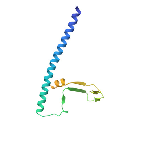
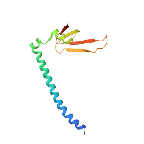
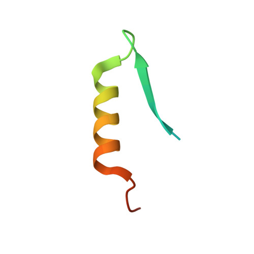
Conservation and divergence of C-terminal domain structure in the retinoblastoma protein family.

Liban, T.J.Medina, E.M.Tripathi, S.Sengupta, S.Henry, R.W.Buchler, N.E.Rubin, S.M.

(2017) Proc Natl Acad Sci U S A 114: 4942-4947

  • DOI: https://doi.org/10.1073/pnas.1619170114
  • Primary Citation of Related Structures:  
    5TUU, 5TUV

  • PubMed Abstract: 

    The retinoblastoma protein (Rb) and the homologous pocket proteins p107 and p130 negatively regulate cell proliferation by binding and inhibiting members of the E2F transcription factor family. The structural features that distinguish Rb from other pocket proteins have been unclear but are critical for understanding their functional diversity and determining why Rb has unique tumor suppressor activities. We describe here important differences in how the Rb and p107 C-terminal domains (CTDs) associate with the coiled-coil and marked-box domains (CMs) of E2Fs. We find that although CTD-CM binding is conserved across protein families, Rb and p107 CTDs show clear preferences for different E2Fs. A crystal structure of the p107 CTD bound to E2F5 and its dimer partner DP1 reveals the molecular basis for pocket protein-E2F binding specificity and how cyclin-dependent kinases differentially regulate pocket proteins through CTD phosphorylation. Our structural and biochemical data together with phylogenetic analyses of Rb and E2F proteins support the conclusion that Rb evolved specific structural motifs that confer its unique capacity to bind with high affinity those E2Fs that are the most potent activators of the cell cycle.


  • Organizational Affiliation

    Department of Chemistry and Biochemistry, University of California, Santa Cruz, CA 95064.


Macromolecules
Find similar proteins by:  (by identity cutoff)  |  3D Structure
Entity ID: 1
MoleculeChains Sequence LengthOrganismDetailsImage
Transcription factor DP1
A, D
155Homo sapiensMutation(s): 0 
Gene Names: TFDP1DP1
UniProt & NIH Common Fund Data Resources
Find proteins for Q14186 (Homo sapiens)
Explore Q14186 
Go to UniProtKB:  Q14186
PHAROS:  Q14186
GTEx:  ENSG00000198176 
Entity Groups  
Sequence Clusters30% Identity50% Identity70% Identity90% Identity95% Identity100% Identity
UniProt GroupQ14186
Sequence Annotations
Expand
  • Reference Sequence
Find similar proteins by:  (by identity cutoff)  |  3D Structure
Entity ID: 2
MoleculeChains Sequence LengthOrganismDetailsImage
Transcription factor E2F5
B, E
112Homo sapiensMutation(s): 0 
Gene Names: E2F5
UniProt & NIH Common Fund Data Resources
Find proteins for Q15329 (Homo sapiens)
Explore Q15329 
Go to UniProtKB:  Q15329
PHAROS:  Q15329
GTEx:  ENSG00000133740 
Entity Groups  
Sequence Clusters30% Identity50% Identity70% Identity90% Identity95% Identity100% Identity
UniProt GroupQ15329
Sequence Annotations
Expand
  • Reference Sequence
Find similar proteins by:  (by identity cutoff)  |  3D Structure
Entity ID: 3
MoleculeChains Sequence LengthOrganismDetailsImage
Retinoblastoma-like protein 1
C, F
41Homo sapiensMutation(s): 0 
Gene Names: RBL1
UniProt & NIH Common Fund Data Resources
Find proteins for P28749 (Homo sapiens)
Explore P28749 
Go to UniProtKB:  P28749
PHAROS:  P28749
GTEx:  ENSG00000080839 
Entity Groups  
Sequence Clusters30% Identity50% Identity70% Identity90% Identity95% Identity100% Identity
UniProt GroupP28749
Sequence Annotations
Expand
  • Reference Sequence
Experimental Data & Validation

Experimental Data

  • Method: X-RAY DIFFRACTION
  • Resolution: 2.90 Å
  • R-Value Free: 0.260 
  • R-Value Work: 0.212 
  • R-Value Observed: 0.215 
  • Space Group: P 1 21 1
Unit Cell:
Length ( Å )Angle ( ˚ )
a = 60.98α = 90
b = 57.34β = 96.64
c = 99.198γ = 90
Software Package:
Software NamePurpose
PHENIXrefinement
iMOSFLMdata reduction
SCALAdata scaling
AutoSolphasing

Structure Validation

View Full Validation Report


Entry History & Funding Information

Deposition Data


Funding OrganizationLocationGrant Number
National Institutes of Health/National Cancer Institute (NIH/NCI)United StatesR01CA132685

Revision History  (Full details and data files)

  • Version 1.0: 2017-05-03
    Type: Initial release
  • Version 1.1: 2017-05-10
    Changes: Database references
  • Version 1.2: 2017-05-24
    Changes: Database references
  • Version 1.3: 2017-09-27
    Changes: Author supporting evidence
  • Version 1.4: 2019-12-04
    Changes: Author supporting evidence
  • Version 1.5: 2024-03-06
    Changes: Data collection, Database references