5TTJ

Crystal Structure of Nicotine Oxidoreductase from Pseudomonas putida


Experimental Data Snapshot

  • Method: X-RAY DIFFRACTION
  • Resolution: 2.20 Å
  • R-Value Free: 0.231 
  • R-Value Work: 0.175 
  • R-Value Observed: 0.176 

wwPDB Validation   3D Report Full Report


Ligand Structure Quality Assessment 


This is version 1.4 of the entry. See complete history


Literature

Structural Analysis Provides Mechanistic Insight into Nicotine Oxidoreductase from Pseudomonas putida.

Tararina, M.A.Janda, K.D.Allen, K.N.

(2016) Biochemistry 55: 6595-6598

  • DOI: https://doi.org/10.1021/acs.biochem.6b00963
  • Primary Citation of Related Structures:  
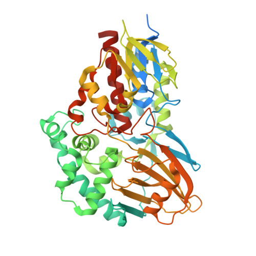
    5TJR, 5TTJ, 5TTK

  • PubMed Abstract: 

    The first structure of nicotine oxidoreductase (NicA2) was determined by X-ray crystallography. Pseudomonas putida has evolved nicotine-degrading activity to provide a source of carbon and nitrogen. The structure establishes NicA2 as a member of the monoamine oxidase family. Residues 1-50 are disordered and may play a role in localization. The nicotine-binding site proximal to the isoalloxazine ring of flavin shows an unusual composition of the classical aromatic cage (W427 and N462). The active site architecture is consistent with the proposed binding of the deprotonated form of the substrate and the flavin-dependent oxidation of the pyrrolidone C-N bond followed by nonenzymatic hydrolysis.


  • Organizational Affiliation

    Program in Biomolecular Pharmacology, Boston University School of Medicine , 72 East Concord Street, Boston, Massachusetts 02118, United States.


Macromolecules
Find similar proteins by:  (by identity cutoff)  |  3D Structure
Entity ID: 1
MoleculeChains Sequence LengthOrganismDetailsImage
Amine oxidase
A, B
490Pseudomonas putida S16Mutation(s): 0 
Gene Names: PPS_4081
UniProt
Find proteins for F8G0P2 (Pseudomonas putida (strain DSM 28022 / S16))
Explore F8G0P2 
Go to UniProtKB:  F8G0P2
Entity Groups  
Sequence Clusters30% Identity50% Identity70% Identity90% Identity95% Identity100% Identity
UniProt GroupF8G0P2
Sequence Annotations
Expand
  • Reference Sequence
Small Molecules
Ligands 1 Unique
IDChains Name / Formula / InChI Key2D Diagram3D Interactions
FAD
Query on FAD

Download Ideal Coordinates CCD File 
C [auth A],
D [auth B]
FLAVIN-ADENINE DINUCLEOTIDE
C27 H33 N9 O15 P2
VWWQXMAJTJZDQX-UYBVJOGSSA-N
Experimental Data & Validation

Experimental Data

  • Method: X-RAY DIFFRACTION
  • Resolution: 2.20 Å
  • R-Value Free: 0.231 
  • R-Value Work: 0.175 
  • R-Value Observed: 0.176 
  • Space Group: P 1 21 1
Unit Cell:
Length ( Å )Angle ( ˚ )
a = 67.973α = 90
b = 61.564β = 92.51
c = 92.277γ = 90
Software Package:
Software NamePurpose
PHENIXrefinement
XDSdata scaling
PHENIXphasing
PHENIXmodel building

Structure Validation

View Full Validation Report


Ligand Structure Quality Assessment 


Entry History & Funding Information

Deposition Data


Funding OrganizationLocationGrant Number
National Institutes of Health/National Institute on Drug Abuse (NIH/NIDA)United StatesDA041839
National Institutes of Health/National Institute of General Medical Sciences (NIH/NIGMS)United StatesT32GM008541

Revision History  (Full details and data files)

  • Version 1.0: 2017-02-08
    Type: Initial release
  • Version 1.1: 2017-09-27
    Changes: Author supporting evidence, Refinement description
  • Version 1.2: 2018-04-18
    Changes: Data collection, Database references
  • Version 1.3: 2019-12-11
    Changes: Advisory, Author supporting evidence, Derived calculations
  • Version 1.4: 2024-03-06
    Changes: Data collection, Database references