


A 1.85A X-Ray Structure from Peptoclostridium difficile 630 of a Hypothetical Protein
Brunzelle, J.S., Minasov, G., Anderson, W.F., Center for Structural Genomics of Infectious Diseases (CSGID)To be published.
Experimental Data Snapshot
wwPDB Validation 3D Report Full Report

Entity ID: 1 | |||||
|---|---|---|---|---|---|
| Molecule | Chains | Sequence Length | Organism | Details | Image |
| Putative exported protein | 248 | Clostridioides difficile 630 | Mutation(s): 0 Gene Names: CD630_21270 | 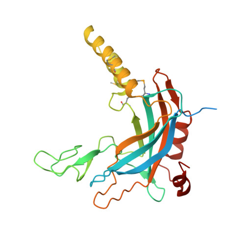 | |
UniProt | |||||
Find proteins for Q185R5 (Clostridioides difficile (strain 630)) Explore Q185R5 Go to UniProtKB: Q185R5 | |||||
Entity Groups | |||||
| Sequence Clusters | 30% Identity50% Identity70% Identity90% Identity95% Identity100% Identity | ||||
| UniProt Group | Q185R5 | ||||
Sequence AnnotationsExpand | |||||
| |||||
| Modified Residues 1 Unique | |||||
|---|---|---|---|---|---|
| ID | Chains | Type | Formula | 2D Diagram | Parent |
| MSE Query on MSE | A, B | L-PEPTIDE LINKING | C5 H11 N O2 Se |  | MET |
| Length ( Å ) | Angle ( ˚ ) |
|---|---|
| a = 30.78 | α = 73.38 |
| b = 51.04 | β = 82.06 |
| c = 81.12 | γ = 86.35 |
| Software Name | Purpose |
|---|---|
| PHENIX | refinement |
| XDS | data reduction |
| Aimless | data scaling |
| PHENIX | phasing |
RCSB PDB (citation) is hosted by
RCSB PDB is a member of the

