5TSH

PilB from Geobacter metallireducens bound to AMP-PNP


Experimental Data Snapshot

  • Method: X-RAY DIFFRACTION
  • Resolution: 2.30 Å
  • R-Value Free: 0.218 
  • R-Value Work: 0.184 
  • R-Value Observed: 0.184 

wwPDB Validation   3D Report Full Report


Ligand Structure Quality Assessment 


This is version 1.3 of the entry. See complete history


Literature

The molecular mechanism of the type IVa pilus motors.

McCallum, M.Tammam, S.Khan, A.Burrows, L.L.Howell, P.L.

(2017) Nat Commun 8: 15091-15091

  • DOI: https://doi.org/10.1038/ncomms15091
  • Primary Citation of Related Structures:  
    5TSG, 5TSH

  • PubMed Abstract: 

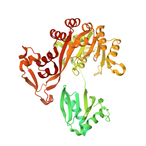
    Type IVa pili are protein filaments essential for virulence in many bacterial pathogens; they extend and retract from the surface of bacterial cells to pull the bacteria forward. The motor ATPase PilB powers pilus assembly. Here we report the structures of the core ATPase domains of Geobacter metallireducens PilB bound to ADP and the non-hydrolysable ATP analogue, AMP-PNP, at 3.4 and 2.3 Å resolution, respectively. These structures reveal important differences in nucleotide binding between chains. Analysis of these differences reveals the sequential turnover of nucleotide, and the corresponding domain movements. Our data suggest a clockwise rotation of the central sub-pores of PilB, which through interactions with PilC, would support the assembly of a right-handed helical pilus. Our analysis also suggests a counterclockwise rotation of the C2 symmetric PilT that would enable right-handed pilus disassembly. The proposed model provides insight into how this family of ATPases can power pilus extension and retraction.


  • Organizational Affiliation

    Department of Biochemistry, University of Toronto, Toronto, Ontario, Canada M5S 1A8.


Macromolecules
Find similar proteins by:  (by identity cutoff)  |  3D Structure
Entity ID: 1
MoleculeChains Sequence LengthOrganismDetailsImage
Type IV pilus biogenesis ATPase PilB
A, B, C, D, E
A, B, C, D, E, F
588Geobacter metallireducens GS-15Mutation(s): 0 
Gene Names: pilBGmet_1393
UniProt
Find proteins for Q39VU7 (Geobacter metallireducens (strain ATCC 53774 / DSM 7210 / GS-15))
Explore Q39VU7 
Go to UniProtKB:  Q39VU7
Entity Groups  
Sequence Clusters30% Identity50% Identity70% Identity90% Identity95% Identity100% Identity
UniProt GroupQ39VU7
Sequence Annotations
Expand
  • Reference Sequence
Small Molecules
Ligands 4 Unique
IDChains Name / Formula / InChI Key2D Diagram3D Interactions
ANP
Query on ANP

Download Ideal Coordinates CCD File 
H [auth A],
K [auth B],
M [auth C],
P [auth D]
PHOSPHOAMINOPHOSPHONIC ACID-ADENYLATE ESTER
C10 H17 N6 O12 P3
PVKSNHVPLWYQGJ-KQYNXXCUSA-N
ADP
Query on ADP

Download Ideal Coordinates CCD File 
R [auth E],
T [auth F]
ADENOSINE-5'-DIPHOSPHATE
C10 H15 N5 O10 P2
XTWYTFMLZFPYCI-KQYNXXCUSA-N
ZN
Query on ZN

Download Ideal Coordinates CCD File 
G [auth A]
J [auth B]
L [auth C]
O [auth D]
Q [auth E]
G [auth A],
J [auth B],
L [auth C],
O [auth D],
Q [auth E],
S [auth F]
ZINC ION
Zn
PTFCDOFLOPIGGS-UHFFFAOYSA-N
MG
Query on MG

Download Ideal Coordinates CCD File 
I [auth A],
N [auth C]
MAGNESIUM ION
Mg
JLVVSXFLKOJNIY-UHFFFAOYSA-N
Experimental Data & Validation

Experimental Data

Unit Cell:
Length ( Å )Angle ( ˚ )
a = 74.32α = 112.99
b = 99.96β = 106.6
c = 109.79γ = 88.91
Software Package:
Software NamePurpose
PHENIXrefinement
XDSdata reduction
XDSdata scaling
PHENIXphasing

Structure Validation

View Full Validation Report


Ligand Structure Quality Assessment 


Entry History & Funding Information

Deposition Data


Funding OrganizationLocationGrant Number
Canadian Institutes of Health Research (CIHR)CanadaMOP 93585

Revision History  (Full details and data files)

  • Version 1.0: 2017-05-17
    Type: Initial release
  • Version 1.1: 2017-09-06
    Changes: Author supporting evidence
  • Version 1.2: 2020-01-08
    Changes: Author supporting evidence
  • Version 1.3: 2023-10-04
    Changes: Data collection, Database references, Derived calculations, Refinement description