5TQS

Phospholipase C gamma-1 C-terminal SH2 domain bound to a phosphopeptide derived from the receptor tyrosine kinase ErbB2


Experimental Data Snapshot

  • Method: X-RAY DIFFRACTION
  • Resolution: 1.88 Å
  • R-Value Free: 0.237 
  • R-Value Work: 0.189 
  • R-Value Observed: 0.191 

wwPDB Validation   3D Report Full Report


This is version 1.6 of the entry. See complete history


Literature

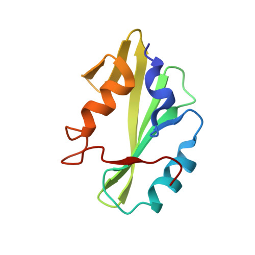
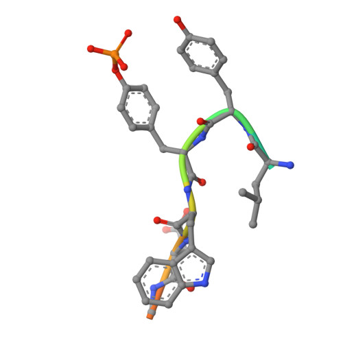
Multimodal Recognition of Diverse Peptides by the C-Terminal SH2 Domain of Phospholipase C-gamma 1 Protein.

McKercher, M.A.Guan, X.Tan, Z.Wuttke, D.S.

(2017) Biochemistry 56: 2225-2237

  • DOI: https://doi.org/10.1021/acs.biochem.7b00023
  • Primary Citation of Related Structures:  
    5TNW, 5TO4, 5TQ1, 5TQS

  • PubMed Abstract: 

    SH2 domains recognize phosphotyrosine (pY)-containing peptide ligands and play key roles in the regulation of receptor tyrosine kinase pathways. Each SH2 domain has individualized specificity, encoded in the amino acids neighboring the pY, for defined targets that convey their distinct functions. The C-terminal SH2 domain (PLCC) of the phospholipase C-γ1 full-length protein (PLCγ1) typically binds peptides containing small and hydrophobic amino acids adjacent to the pY, including a peptide derived from platelet-derived growth factor receptor B (PDGFRB) and an intraprotein recognition site (Y783 of PLCγ1) involved in the regulation of the protein's lipase activity. Remarkably, PLCC also recognizes unexpected peptides containing amino acids with polar or bulky side chains that deviate from this pattern. This versatility in recognition specificity may allow PLCγ1 to participate in diverse, previously unrecognized, signaling pathways in response to binding chemically dissimilar partners. We have used structural approaches, including nuclear magnetic resonance and X-ray crystallography, to elucidate the mechanisms of noncognate peptide binding to PLCC by ligands derived from receptor tyrosine kinase ErbB2 and from the insulin receptor. The high-resolution peptide-bound structures reveal that PLCC has a relatively static backbone but contains a chemically rich protein surface comprised of a combination of hydrophobic pockets and amino acids with charged side chains. We demonstrate that this expansive and chemically diverse PLCC interface, in addition to peptide conformational plasticity, permits PLCC to recognize specific noncognate peptide ligands with multimodal specificity.


  • Organizational Affiliation

    Department of Chemistry and Biochemistry, University of Colorado Boulder , Boulder, Colorado 80309, United States.


Macromolecules
Find similar proteins by:  (by identity cutoff)  |  3D Structure
Entity ID: 1
MoleculeChains Sequence LengthOrganismDetailsImage
1-phosphatidylinositol 4,5-bisphosphate phosphodiesterase gamma-1
A, B, C, D
101Bos taurusMutation(s): 0 
Gene Names: PLCG1
EC: 3.1.4.11
UniProt
Find proteins for P08487 (Bos taurus)
Explore P08487 
Go to UniProtKB:  P08487
Entity Groups  
Sequence Clusters30% Identity50% Identity70% Identity90% Identity95% Identity100% Identity
UniProt GroupP08487
Sequence Annotations
Expand
  • Reference Sequence

Find similar proteins by:  Sequence   |   3D Structure  

Entity ID: 2
MoleculeChains Sequence LengthOrganismDetailsImage
Receptor protein-tyrosine kinaseE,
F,
G [auth H],
H [auth G]
11Homo sapiensMutation(s): 0 
UniProt & NIH Common Fund Data Resources
Find proteins for P04626 (Homo sapiens)
Explore P04626 
Go to UniProtKB:  P04626
PHAROS:  P04626
GTEx:  ENSG00000141736 
Entity Groups  
Sequence Clusters30% Identity50% Identity70% Identity90% Identity95% Identity100% Identity
UniProt GroupP04626
Sequence Annotations
Expand
  • Reference Sequence
Small Molecules
Modified Residues  1 Unique
IDChains TypeFormula2D DiagramParent
PTR
Query on PTR
E,
F,
G [auth H],
H [auth G]
L-PEPTIDE LINKINGC9 H12 N O6 PTYR
Experimental Data & Validation

Experimental Data

  • Method: X-RAY DIFFRACTION
  • Resolution: 1.88 Å
  • R-Value Free: 0.237 
  • R-Value Work: 0.189 
  • R-Value Observed: 0.191 
  • Space Group: P 1 21 1
Unit Cell:
Length ( Å )Angle ( ˚ )
a = 65.32α = 90
b = 30.62β = 100.12
c = 104.57γ = 90
Software Package:
Software NamePurpose
SCALAdata scaling
PHASERphasing
PHENIXrefinement
PDB_EXTRACTdata extraction
MOSFLMdata reduction

Structure Validation

View Full Validation Report


Entry History & Funding Information

Deposition Data


Funding OrganizationLocationGrant Number
National Science Foundation (NSF, United States)United StatesMCB1121842

Revision History  (Full details and data files)

  • Version 1.0: 2017-04-19
    Type: Initial release
  • Version 1.1: 2017-04-26
    Changes: Database references
  • Version 1.2: 2017-05-10
    Changes: Database references
  • Version 1.3: 2017-09-27
    Changes: Author supporting evidence
  • Version 1.4: 2019-11-27
    Changes: Author supporting evidence
  • Version 1.5: 2023-10-04
    Changes: Data collection, Database references, Refinement description
  • Version 1.6: 2023-11-15
    Changes: Data collection