5TOI

Crystal Structure of the Marburg Virus VP35 Oligomerization Domain P4222


Experimental Data Snapshot

  • Method: X-RAY DIFFRACTION
  • Resolution: 2.19 Å
  • R-Value Free: 0.342 
  • R-Value Work: 0.312 
  • R-Value Observed: 0.315 

wwPDB Validation   3D Report Full Report


This is version 1.6 of the entry. See complete history


Literature

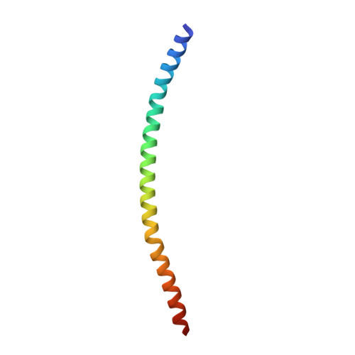
Crystal Structure of the Marburg Virus VP35 Oligomerization Domain.

Bruhn, J.F.Kirchdoerfer, R.N.Urata, S.M.Li, S.Tickle, I.J.Bricogne, G.Saphire, E.O.

(2017) J Virol 91

  • DOI: https://doi.org/10.1128/JVI.01085-16
  • Primary Citation of Related Structures:  
    5TOH, 5TOI

  • PubMed Abstract: 

    Marburg virus (MARV) is a highly pathogenic filovirus that is classified in a genus distinct from that of Ebola virus (EBOV) (genera Marburgvirus and Ebolavirus, respectively). Both viruses produce a multifunctional protein termed VP35, which acts as a polymerase cofactor, a viral protein chaperone, and an antagonist of the innate immune response. VP35 contains a central oligomerization domain with a predicted coiled-coil motif. This domain has been shown to be essential for RNA polymerase function. Here we present crystal structures of the MARV VP35 oligomerization domain. These structures and accompanying biophysical characterization suggest that MARV VP35 is a trimer. In contrast, EBOV VP35 is likely a tetramer in solution. Differences in the oligomeric state of this protein may explain mechanistic differences in replication and immune evasion observed for MARV and EBOV.


  • Organizational Affiliation

    Department of Immunology and Microbial Science, The Scripps Research Institute, La Jolla, California, USA.


Macromolecules
Find similar proteins by:  (by identity cutoff)  |  3D Structure
Entity ID: 1
MoleculeChains Sequence LengthOrganismDetailsImage
Polymerase cofactor VP35
A, B, C
72Marburg virus - Musoke, Kenya, 1980Mutation(s): 0 
Gene Names: VP35
UniProt
Find proteins for P35259 (Lake Victoria marburgvirus (strain Musoke-80))
Explore P35259 
Go to UniProtKB:  P35259
Entity Groups  
Sequence Clusters30% Identity50% Identity70% Identity90% Identity95% Identity100% Identity
UniProt GroupP35259
Sequence Annotations
Expand
  • Reference Sequence
Experimental Data & Validation

Experimental Data

  • Method: X-RAY DIFFRACTION
  • Resolution: 2.19 Å
  • R-Value Free: 0.342 
  • R-Value Work: 0.312 
  • R-Value Observed: 0.315 
  • Space Group: P 42 2 2
Unit Cell:
Length ( Å )Angle ( ˚ )
a = 46.49α = 90
b = 46.49β = 90
c = 310.93γ = 90
Software Package:
Software NamePurpose
PDB_EXTRACTdata extraction
Aimlessdata scaling
XDSdata reduction
PHASERphasing
STARANISOdata scaling
BUSTERrefinement

Structure Validation

View Full Validation Report


Entry History & Funding Information

Deposition Data


Funding OrganizationLocationGrant Number
National Institutes of Health/National Institute Of Allergy and Infectious Diseases (NIH/NIAID)United StatesT32 AI007606
National Institutes of Health/National Institute Of Allergy and Infectious Diseases (NIH/NIAID)United StatesR01 AI118016
Burroughs Wellcome FundUnited States--
Department of Energy (DOE, United States)United StatesDE-AC02-76SF00515
National Institutes of Health/National Institute of General Medical Sciences (NIH/NIGMS)United StatesP41GM103393

Revision History  (Full details and data files)

  • Version 1.0: 2016-11-09
    Type: Initial release
  • Version 1.1: 2016-11-30
    Changes: Database references
  • Version 1.2: 2017-01-11
    Changes: Database references
  • Version 1.3: 2017-02-08
    Changes: Database references
  • Version 1.4: 2017-09-27
    Changes: Author supporting evidence, Refinement description
  • Version 1.5: 2019-12-04
    Changes: Author supporting evidence
  • Version 1.6: 2023-10-04
    Changes: Data collection, Database references, Refinement description