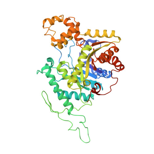
5TLC

Crystal structure of BdsA from Bacillus subtilis WU-S2B


Experimental Data Snapshot

  • Method: X-RAY DIFFRACTION
  • Resolution: 2.80 Å
  • R-Value Free: 0.274 
  • R-Value Work: 0.212 
  • R-Value Observed: 0.215 

wwPDB Validation   3D Report Full Report


This is version 1.2 of the entry. See complete history


Literature

Crystal structure of dibenzothiophene sulfone monooxygenase BdsA from Bacillus subtilis WU-S2B

Okai, M.Lee, W.C.Guan, L.J.Ohshiro, T.Izumi, Y.Tanokura, M.

(2017) Proteins 85: 1171-1177

  • DOI: https://doi.org/10.1002/prot.25267
  • Primary Citation of Related Structures:  
    5TLC

  • PubMed Abstract: 

    The dibenzothiophene (DBT) sulfone monooxygenase BdsA from Bacillus subtilis WU-S2B catalyzes the conversion of DBT sulfone to 2'-hydroxybiphenyl 2-sulfinate. We report the crystal structures of BdsA at a resolution of 2.80 Å. BdsA exists as a homotetramer with a dimer-of-dimers configuration in the crystal, and the interaction between E288 and R296 in BdsA is important for tetramer formation. A structural comparison with homologous proteins shows that the orientation and location of the α9-α12 helices in BdsA are closer to those of the closed form than those of the open form in the EDTA monooxygenase EmoA. Proteins 2017; 85:1171-1177. © 2017 Wiley Periodicals, Inc.


  • Organizational Affiliation

    Department of Applied Biological Chemistry, Graduate School of Agricultural and Life Sciences, University of Tokyo, Bunkyo-ku, Tokyo, 113-8657, Japan.


Macromolecules
Find similar proteins by:  (by identity cutoff)  |  3D Structure
Entity ID: 1
MoleculeChains Sequence LengthOrganismDetailsImage
Dibenzothiophene desulfurization enzyme A
A, B, C, D
453Bacillus subtilisMutation(s): 0 
Gene Names: bdsA
UniProt
Find proteins for Q8GRC7 (Bacillus subtilis)
Explore Q8GRC7 
Go to UniProtKB:  Q8GRC7
Entity Groups  
Sequence Clusters30% Identity50% Identity70% Identity90% Identity95% Identity100% Identity
UniProt GroupQ8GRC7
Sequence Annotations
Expand
  • Reference Sequence
Experimental Data & Validation

Experimental Data

  • Method: X-RAY DIFFRACTION
  • Resolution: 2.80 Å
  • R-Value Free: 0.274 
  • R-Value Work: 0.212 
  • R-Value Observed: 0.215 
  • Space Group: P 32
Unit Cell:
Length ( Å )Angle ( ˚ )
a = 84.126α = 90
b = 84.126β = 90
c = 269.456γ = 120
Software Package:
Software NamePurpose
REFMACrefinement
XDSdata reduction
SCALAdata scaling

Structure Validation

View Full Validation Report


Entry History 

Deposition Data

Revision History  (Full details and data files)

  • Version 1.0: 2017-05-03
    Type: Initial release
  • Version 1.1: 2017-05-31
    Changes: Database references
  • Version 1.2: 2024-03-20
    Changes: Data collection, Database references