5TL4

Crystal structure of Sphingomonas paucimobilis aryl O-demethylase LigM


Experimental Data Snapshot

  • Method: X-RAY DIFFRACTION
  • Resolution: 1.75 Å
  • R-Value Free: 0.212 
  • R-Value Work: 0.170 
  • R-Value Observed: 0.170 

wwPDB Validation   3D Report Full Report


This is version 1.5 of the entry. See complete history


Literature

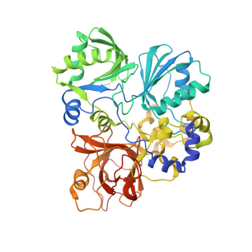
Structure of aryl O-demethylase offers molecular insight into a catalytic tyrosine-dependent mechanism.

Kohler, A.C.Mills, M.J.L.Adams, P.D.Simmons, B.A.Sale, K.L.

(2017) Proc Natl Acad Sci U S A 114: E3205-E3214

  • DOI: https://doi.org/10.1073/pnas.1619263114
  • Primary Citation of Related Structures:  
    5TL4

  • PubMed Abstract: 

    Some strains of soil and marine bacteria have evolved intricate metabolic pathways for using environmentally derived aromatics as a carbon source. Many of these metabolic pathways go through intermediates such as vanillate, 3- O -methylgallate, and syringate. Demethylation of these compounds is essential for downstream aryl modification, ring opening, and subsequent assimilation of these compounds into the tricarboxylic acid (TCA) cycle, and, correspondingly, there are a variety of associated aryl demethylase systems that vary in complexity. Intriguingly, only a basic understanding of the least complex system, the tetrahydrofolate-dependent aryl demethylase LigM from Sphingomonas paucimobilis , a bacterial strain that metabolizes lignin-derived aromatics, was previously available. LigM-catalyzed demethylation enables further modification and ring opening of the single-ring aromatics vanillate and 3- O -methylgallate, which are common byproducts of biofuel production. Here, we characterize aryl O -demethylation by LigM and report its 1.81-Å crystal structure, revealing a unique demethylase fold and a canonical folate-binding domain. Structural homology and geometry optimization calculations enabled the identification of LigM's tetrahydrofolate-binding site and protein-folate interactions. Computationally guided mutagenesis and kinetic analyses allowed the identification of the enzyme's aryl-binding site location and determination of its unique, catalytic tyrosine-dependent reaction mechanism. This work defines LigM as a distinct demethylase, both structurally and functionally, and provides insight into demethylation and its reaction requirements. These results afford the mechanistic details required for efficient utilization of LigM as a tool for aryl O -demethylation and as a component of synthetic biology efforts to valorize previously underused aromatic compounds.


  • Organizational Affiliation

    Joint BioEnergy Institute, Emeryville, CA 94608.


Macromolecules
Find similar proteins by:  (by identity cutoff)  |  3D Structure
Entity ID: 1
MoleculeChains Sequence LengthOrganismDetailsImage
Vanillate/3-O-methylgallate O-demethylase
A, B, C, D
475Sphingomonas paucimobilisMutation(s): 0 
Gene Names: ligM
UniProt
Find proteins for G2IQS7 (Sphingobium sp. (strain NBRC 103272 / SYK-6))
Explore G2IQS7 
Go to UniProtKB:  G2IQS7
Entity Groups  
Sequence Clusters30% Identity50% Identity70% Identity90% Identity95% Identity100% Identity
UniProt GroupG2IQS7
Sequence Annotations
Expand
  • Reference Sequence
Small Molecules
Ligands 4 Unique
IDChains Name / Formula / InChI Key2D Diagram3D Interactions
GOL
Query on GOL

Download Ideal Coordinates CCD File 
H [auth A],
J [auth B],
N [auth C],
R [auth D]
GLYCEROL
C3 H8 O3
PEDCQBHIVMGVHV-UHFFFAOYSA-N
CL
Query on CL

Download Ideal Coordinates CCD File 
E [auth A],
I [auth B],
K [auth C],
L [auth C],
O [auth D]
CHLORIDE ION
Cl
VEXZGXHMUGYJMC-UHFFFAOYSA-M
NA
Query on NA

Download Ideal Coordinates CCD File 
F [auth A],
M [auth C],
P [auth D],
Q [auth D]
SODIUM ION
Na
FKNQFGJONOIPTF-UHFFFAOYSA-N
NH4
Query on NH4

Download Ideal Coordinates CCD File 
G [auth A]AMMONIUM ION
H4 N
QGZKDVFQNNGYKY-UHFFFAOYSA-O
Experimental Data & Validation

Experimental Data

  • Method: X-RAY DIFFRACTION
  • Resolution: 1.75 Å
  • R-Value Free: 0.212 
  • R-Value Work: 0.170 
  • R-Value Observed: 0.170 
  • Space Group: P 21 21 21
Unit Cell:
Length ( Å )Angle ( ˚ )
a = 112.837α = 90
b = 126.184β = 90
c = 155.646γ = 90
Software Package:
Software NamePurpose
PHENIXrefinement
HKL-2000data reduction
HKL-2000data scaling
PHENIXphasing

Structure Validation

View Full Validation Report


Entry History & Funding Information

Deposition Data


Funding OrganizationLocationGrant Number
Department of Energy (DOE, United States)United StatesDE-AC02-05CH11231

Revision History  (Full details and data files)

  • Version 1.0: 2017-04-12
    Type: Initial release
  • Version 1.1: 2017-04-19
    Changes: Database references
  • Version 1.2: 2017-05-03
    Changes: Database references
  • Version 1.3: 2017-09-27
    Changes: Author supporting evidence
  • Version 1.4: 2019-12-04
    Changes: Author supporting evidence
  • Version 1.5: 2024-03-06
    Changes: Data collection, Database references, Derived calculations