5TKA

Structure of the HD-domain phosphohydrolase OxsA


Experimental Data Snapshot

  • Method: X-RAY DIFFRACTION
  • Resolution: 2.05 Å
  • R-Value Free: 0.254 
  • R-Value Work: 0.206 
  • R-Value Observed: 0.209 

wwPDB Validation   3D Report Full Report


This is version 1.5 of the entry. See complete history


Literature

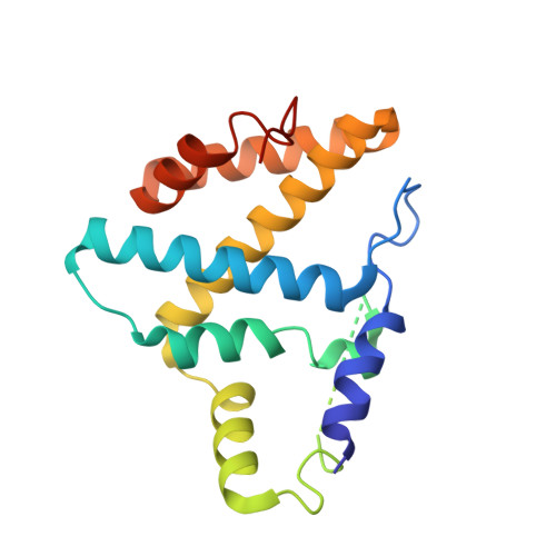
An HD domain phosphohydrolase active site tailored for oxetanocin-A biosynthesis.

Bridwell-Rabb, J.Kang, G.Zhong, A.Liu, H.W.Drennan, C.L.

(2016) Proc Natl Acad Sci U S A 113: 13750-13755

  • DOI: https://doi.org/10.1073/pnas.1613610113
  • Primary Citation of Related Structures:  
    5TK6, 5TK7, 5TK8, 5TK9, 5TKA

  • PubMed Abstract: 

    HD domain phosphohydrolase enzymes are characterized by a conserved set of histidine and aspartate residues that coordinate an active site metallocenter. Despite the important roles these enzymes play in nucleotide metabolism and signal transduction, few have been both biochemically and structurally characterized. Here, we present X-ray crystal structures and biochemical characterization of the Bacillus megaterium HD domain phosphohydrolase OxsA, involved in the biosynthesis of the antitumor, antiviral, and antibacterial compound oxetanocin-A. These studies reveal a previously uncharacterized reaction for this family; OxsA catalyzes the conversion of a triphosphorylated compound into a nucleoside, releasing one molecule of inorganic phosphate at a time. Remarkably, this functionality is a result of the OxsA active site, which based on structural and kinetic analyses has been tailored to bind the small, four-membered ring of oxetanocin-A over larger substrates. Furthermore, our OxsA structures show an active site that switches from a dinuclear to a mononuclear metal center as phosphates are eliminated from substrate.


  • Organizational Affiliation

    Howard Hughes Medical Institute, Massachusetts Institute of Technology, Cambridge, MA 02139.


Macromolecules
Find similar proteins by:  (by identity cutoff)  |  3D Structure
Entity ID: 1
MoleculeChains Sequence LengthOrganismDetailsImage
OxsA protein194Priestia megateriumMutation(s): 0 
Gene Names: oxsA
UniProt
Find proteins for O24769 (Priestia megaterium)
Explore O24769 
Go to UniProtKB:  O24769
Entity Groups  
Sequence Clusters30% Identity50% Identity70% Identity90% Identity95% Identity100% Identity
UniProt GroupO24769
Sequence Annotations
Expand
  • Reference Sequence
Small Molecules
Experimental Data & Validation

Experimental Data

  • Method: X-RAY DIFFRACTION
  • Resolution: 2.05 Å
  • R-Value Free: 0.254 
  • R-Value Work: 0.206 
  • R-Value Observed: 0.209 
  • Space Group: H 3 2
Unit Cell:
Length ( Å )Angle ( ˚ )
a = 143.031α = 90
b = 143.031β = 90
c = 53.786γ = 120
Software Package:
Software NamePurpose
PHENIXrefinement
HKL-2000data reduction
HKL-2000data scaling
PHENIXphasing

Structure Validation

View Full Validation Report


Entry History & Funding Information

Deposition Data


Funding OrganizationLocationGrant Number
National Institutes of Health/National Institute of General Medical Sciences (NIH/NIGMS)United StatesF32-GM108189

Revision History  (Full details and data files)

  • Version 1.0: 2016-11-16
    Type: Initial release
  • Version 1.1: 2016-11-30
    Changes: Database references
  • Version 1.2: 2016-12-14
    Changes: Database references
  • Version 1.3: 2017-09-27
    Changes: Author supporting evidence
  • Version 1.4: 2019-12-25
    Changes: Author supporting evidence
  • Version 1.5: 2023-10-04
    Changes: Data collection, Database references, Derived calculations, Refinement description