Mechanistic and Evolutionary Insights from Comparative Enzymology of Phosphomonoesterases and Phosphodiesterases across the Alkaline Phosphatase Superfamily.
Sunden, F., AlSadhan, I., Lyubimov, A.Y., Ressl, S., Wiersma-Koch, H., Borland, J., Brown, C.L., Johnson, T.A., Singh, Z., Herschlag, D.(2016) J Am Chem Soc 138: 14273-14287
- PubMed: 27670607
- DOI: https://doi.org/10.1021/jacs.6b06186
- Primary Citation of Related Structures:
5TJ3 - PubMed Abstract:
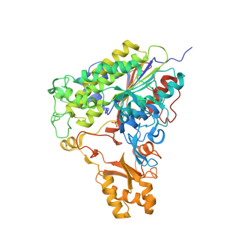
Naively one might have expected an early division between phosphate monoesterases and diesterases of the alkaline phosphatase (AP) superfamily. On the contrary, prior results and our structural and biochemical analyses of phosphate monoesterase PafA, from Chryseobacterium meningosepticum, indicate similarities to a superfamily phosphate diesterase [Xanthomonas citri nucleotide pyrophosphatase/phosphodiesterase (NPP)] and distinct differences from the three metal ion AP superfamily monoesterase, from Escherichia coli AP (EcAP). We carried out a series of experiments to map out and learn from the differences and similarities between these enzymes. First, we asked why there would be independent instances of monoesterases in the AP superfamily? PafA has a much weaker product inhibition and slightly higher activity relative to EcAP, suggesting that different metabolic evolutionary pressures favored distinct active-site architectures. Next, we addressed the preferential phosphate monoester and diester catalysis of PafA and NPP, respectively. We asked whether the >80% sequence differences throughout these scaffolds provide functional specialization for each enzyme's cognate reaction. In contrast to expectations from this model, PafA and NPP mutants with the common subset of active-site groups embedded in each native scaffold had the same monoesterase:diesterase specificities; thus, the >10 7 -fold difference in native specificities appears to arise from distinct interactions at a single phosphoryl substituent. We also uncovered striking mechanistic similarities between the PafA and EcAP monoesterases, including evidence for ground-state destabilization and functional active-site networks that involve different active-site groups but may play analogous catalytic roles. Discovering common network functions may reveal active-site architectural connections that are critical for function, and identifying regions of functional modularity may facilitate the design of new enzymes from existing promiscuous templates. More generally, comparative enzymology and analysis of catalytic promiscuity can provide mechanistic and evolutionary insights.
Organizational Affiliation:
Department of Biochemistry, Beckman Center, Stanford University , Stanford, California 94305, United States.