5TE8

Crystal structure of the midazolam-bound human CYP3A4

  • Classification: OXIDOREDUCTASE
  • Organism(s): Homo sapiens
  • Expression System: Escherichia coli BL21(DE3)
  • Mutation(s): No 
  • Membrane Protein: Yes  OPM

  • Deposited: 2016-09-20 Released: 2016-12-07 
  • Deposition Author(s): Sevrioukova, I., Poulos, T.
  • Funding Organization(s): National Institutes of Health/National Institute of Environmental Health Sciences (NIH/NIEHS), National Institutes of Health/National Institute of General Medical Sciences (NIH/NIGMS)

Experimental Data Snapshot

  • Method: X-RAY DIFFRACTION
  • Resolution: 2.70 Å
  • R-Value Free: 0.292 
  • R-Value Work: 0.224 
  • R-Value Observed: 0.227 

wwPDB Validation   3D Report Full Report


Ligand Structure Quality Assessment 


This is version 1.5 of the entry. See complete history


Literature

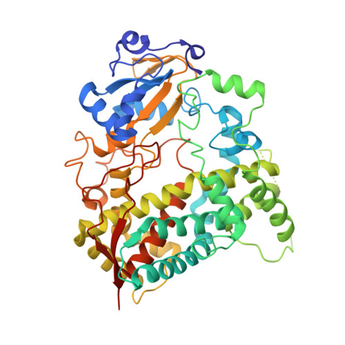
Structural basis for regiospecific midazolam oxidation by human cytochrome P450 3A4.

Sevrioukova, I.F.Poulos, T.L.

(2017) Proc Natl Acad Sci U S A 114: 486-491

  • DOI: https://doi.org/10.1073/pnas.1616198114
  • Primary Citation of Related Structures:  
    5TE8

  • PubMed Abstract: 

    Human cytochrome P450 3A4 (CYP3A4) is a major hepatic and intestinal enzyme that oxidizes more than 60% of administered therapeutics. Knowledge of how CYP3A4 adjusts and reshapes the active site to regioselectively oxidize chemically diverse compounds is critical for better understanding structure-function relations in this important enzyme, improving the outcomes for drug metabolism predictions, and developing pharmaceuticals that have a decreased ability to undergo metabolism and cause detrimental drug-drug interactions. However, there is very limited structural information on CYP3A4-substrate interactions available to date. Despite the vast variety of drugs undergoing metabolism, only the sedative midazolam (MDZ) serves as a marker substrate for the in vivo activity assessment because it is preferentially and regioselectively oxidized by CYP3A4. We solved the 2.7 Å crystal structure of the CYP3A4-MDZ complex, where the drug is well defined and oriented suitably for hydroxylation of the C1 atom, the major site of metabolism. This binding mode requires H-bonding to Ser119 and a dramatic conformational switch in the F-G fragment, which transmits to the adjacent D, E, H, and I helices, resulting in a collapse of the active site cavity and MDZ immobilization. In addition to providing insights on the substrate-triggered active site reshaping (an induced fit), the crystal structure explains the accumulated experimental results, identifies possible effector binding sites, and suggests why MDZ is predominantly metabolized by the CYP3A enzyme subfamily.


  • Organizational Affiliation

    Department of Molecular Biology and Biochemistry, University of California, Irvine, CA 92697-3900; sevrioui@uci.edu.


Macromolecules
Find similar proteins by:  (by identity cutoff)  |  3D Structure
Entity ID: 1
MoleculeChains Sequence LengthOrganismDetailsImage
Cytochrome P450 3A4
A, B, C
487Homo sapiensMutation(s): 0 
Gene Names: CYP3A4CYP3A3
EC: 1.14.13
Membrane Entity: Yes 
UniProt & NIH Common Fund Data Resources
Find proteins for P08684 (Homo sapiens)
Explore P08684 
Go to UniProtKB:  P08684
PHAROS:  P08684
GTEx:  ENSG00000160868 
Entity Groups  
Sequence Clusters30% Identity50% Identity70% Identity90% Identity95% Identity100% Identity
UniProt GroupP08684
Sequence Annotations
Expand
  • Reference Sequence
Small Molecules
Binding Affinity Annotations 
IDSourceBinding Affinity
08J BindingDB:  5TE8 Ki: 2470 (nM) from 1 assay(s)
IC50: min: 8100, max: 3.30e+4 (nM) from 2 assay(s)
Experimental Data & Validation

Experimental Data

  • Method: X-RAY DIFFRACTION
  • Resolution: 2.70 Å
  • R-Value Free: 0.292 
  • R-Value Work: 0.224 
  • R-Value Observed: 0.227 
  • Space Group: P 21 21 21
Unit Cell:
Length ( Å )Angle ( ˚ )
a = 64.289α = 90
b = 117.98β = 90
c = 205.649γ = 90
Software Package:
Software NamePurpose
REFMACrefinement
MOSFLMdata reduction
SCALAdata scaling
PHASERphasing

Structure Validation

View Full Validation Report


Ligand Structure Quality Assessment 


Entry History & Funding Information

Deposition Data


Funding OrganizationLocationGrant Number
National Institutes of Health/National Institute of Environmental Health Sciences (NIH/NIEHS)United StatesES025767
National Institutes of Health/National Institute of General Medical Sciences (NIH/NIGMS)United StatesGM57353

Revision History  (Full details and data files)

  • Version 1.0: 2016-12-07
    Type: Initial release
  • Version 1.1: 2017-01-11
    Changes: Database references
  • Version 1.2: 2017-01-25
    Changes: Database references
  • Version 1.3: 2017-09-20
    Changes: Author supporting evidence
  • Version 1.4: 2019-12-18
    Changes: Author supporting evidence
  • Version 1.5: 2023-10-04
    Changes: Data collection, Database references, Refinement description