5TE3

Crystal structure of Bos taurus opsin at 2.7 Angstrom


Experimental Data Snapshot

  • Method: X-RAY DIFFRACTION
  • Resolution: 2.70 Å
  • R-Value Free: 0.254 
  • R-Value Work: 0.245 
  • R-Value Observed: 0.245 

wwPDB Validation   3D Report Full Report


Ligand Structure Quality Assessment 


This is version 2.1 of the entry. See complete history


Literature

Photocyclic behavior of rhodopsin induced by an atypical isomerization mechanism.

Gulati, S.Jastrzebska, B.Banerjee, S.Placeres, A.L.Miszta, P.Gao, S.Gunderson, K.Tochtrop, G.P.Filipek, S.Katayama, K.Kiser, P.D.Mogi, M.Stewart, P.L.Palczewski, K.

(2017) Proc Natl Acad Sci U S A 114: E2608-E2615

  • DOI: https://doi.org/10.1073/pnas.1617446114
  • Primary Citation of Related Structures:  
    5TE3, 5TE5

  • PubMed Abstract: 

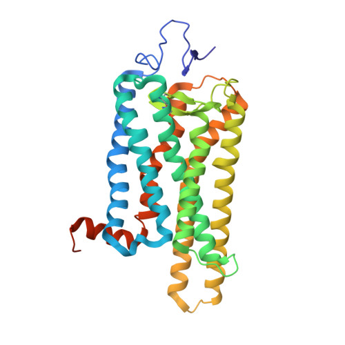
    Vertebrate rhodopsin (Rh) contains 11- cis -retinal as a chromophore to convert light energy into visual signals. On absorption of light, 11- cis -retinal is isomerized to all- trans -retinal, constituting a one-way reaction that activates transducin (G t ) followed by chromophore release. Here we report that bovine Rh, regenerated instead with a six-carbon-ring retinal chromophore featuring a C 11 =C 12 double bond locked in its cis conformation (Rh6mr), employs an atypical isomerization mechanism by converting 11- cis to an 11,13- dicis configuration for prolonged G t activation. Time-dependent UV-vis spectroscopy, HPLC, and molecular mechanics analyses revealed an atypical thermal reisomerization of the 11,13- dicis to the 11- cis configuration on a slow timescale, which enables Rh6mr to function in a photocyclic manner similar to that of microbial Rhs. With this photocyclic behavior, Rh6mr repeatedly recruits and activates G t in response to light stimuli, making it an excellent candidate for optogenetic tools based on retinal analog-bound vertebrate Rhs. Overall, these comprehensive structure-function studies unveil a unique photocyclic mechanism of Rh activation by an 11- cis -to-11,13- dicis isomerization.


  • Organizational Affiliation

    Department of Pharmacology, School of Medicine, Case Western Reserve University, Cleveland, OH 44106.


Macromolecules
Find similar proteins by:  (by identity cutoff)  |  3D Structure
Entity ID: 1
MoleculeChains Sequence LengthOrganismDetailsImage
Rhodopsin348Bos taurusMutation(s): 0 
Membrane Entity: Yes 
UniProt
Find proteins for P02699 (Bos taurus)
Explore P02699 
Go to UniProtKB:  P02699
Entity Groups  
Sequence Clusters30% Identity50% Identity70% Identity90% Identity95% Identity100% Identity
UniProt GroupP02699
Sequence Annotations
Expand
  • Reference Sequence
Oligosaccharides

Help

Entity ID: 2
MoleculeChains Length2D Diagram Glycosylation3D Interactions
alpha-D-mannopyranose-(1-3)-beta-D-mannopyranose-(1-4)-2-acetamido-2-deoxy-beta-D-glucopyranose-(1-4)-2-acetamido-2-deoxy-beta-D-glucopyranose
B
4N-Glycosylation
Glycosylation Resources
GlyTouCan:  G81315DD
GlyCosmos:  G81315DD
GlyGen:  G81315DD
Experimental Data & Validation

Experimental Data

  • Method: X-RAY DIFFRACTION
  • Resolution: 2.70 Å
  • R-Value Free: 0.254 
  • R-Value Work: 0.245 
  • R-Value Observed: 0.245 
  • Space Group: H 3 2
Unit Cell:
Length ( Å )Angle ( ˚ )
a = 242.66α = 90
b = 242.66β = 90
c = 111.77γ = 120
Software Package:
Software NamePurpose
REFMACrefinement
XDSdata reduction
XSCALEdata scaling
REFMACphasing

Structure Validation

View Full Validation Report


Ligand Structure Quality Assessment 


Entry History 

Deposition Data

Revision History  (Full details and data files)

  • Version 1.0: 2017-03-15
    Type: Initial release
  • Version 1.1: 2017-03-29
    Changes: Database references
  • Version 1.2: 2017-04-05
    Changes: Database references
  • Version 2.0: 2020-07-29
    Type: Remediation
    Reason: Carbohydrate remediation
    Changes: Atomic model, Data collection, Derived calculations, Structure summary
  • Version 2.1: 2023-10-04
    Changes: Data collection, Database references, Derived calculations, Refinement description, Structure summary