5T3D

Crystal structure of holo-EntF a nonribosomal peptide synthetase in the thioester-forming conformation


Experimental Data Snapshot

  • Method: X-RAY DIFFRACTION
  • Resolution: 2.80 Å
  • R-Value Free: 0.232 
  • R-Value Work: 0.185 
  • R-Value Observed: 0.188 

wwPDB Validation   3D Report Full Report


Ligand Structure Quality Assessment 


This is version 1.3 of the entry. See complete history


Literature

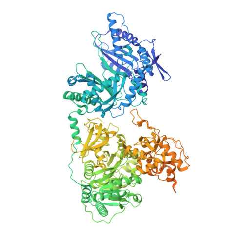
Structures of two distinct conformations of holo-non-ribosomal peptide synthetases.

Drake, E.J.Miller, B.R.Shi, C.Tarrasch, J.T.Sundlov, J.A.Allen, C.L.Skiniotis, G.Aldrich, C.C.Gulick, A.M.

(2016) Nature 529: 235-238

  • DOI: https://doi.org/10.1038/nature16163
  • Primary Citation of Related Structures:  
    4ZXH, 4ZXI, 5T3D

  • PubMed Abstract: 

    Many important natural products are produced by multidomain non-ribosomal peptide synthetases (NRPSs). During synthesis, intermediates are covalently bound to integrated carrier domains and transported to neighbouring catalytic domains in an assembly line fashion. Understanding the structural basis for catalysis with non-ribosomal peptide synthetases will facilitate bioengineering to create novel products. Here we describe the structures of two different holo-non-ribosomal peptide synthetase modules, each revealing a distinct step in the catalytic cycle. One structure depicts the carrier domain cofactor bound to the peptide bond-forming condensation domain, whereas a second structure captures the installation of the amino acid onto the cofactor within the adenylation domain. These structures demonstrate that a conformational change within the adenylation domain guides transfer of intermediates between domains. Furthermore, one structure shows that the condensation and adenylation domains simultaneously adopt their catalytic conformations, increasing the overall efficiency in a revised structural cycle. These structures and the single-particle electron microscopy analysis demonstrate a highly dynamic domain architecture and provide the foundation for understanding the structural mechanisms that could enable engineering of novel non-ribosomal peptide synthetases.


  • Organizational Affiliation

    Hauptman-Woodward Medical Research Institute, 700 Ellicott Street, Buffalo, New York 14203, USA.


Macromolecules
Find similar proteins by:  (by identity cutoff)  |  3D Structure
Entity ID: 1
MoleculeChains Sequence LengthOrganismDetailsImage
Enterobactin synthase component F1,295Escherichia coli K-12Mutation(s): 0 
Gene Names: entFb0586JW0578
EC: 2.7.7
UniProt
Find proteins for P11454 (Escherichia coli (strain K12))
Explore P11454 
Go to UniProtKB:  P11454
Entity Groups  
Sequence Clusters30% Identity50% Identity70% Identity90% Identity95% Identity100% Identity
UniProt GroupP11454
Sequence Annotations
Expand
  • Reference Sequence
Small Molecules
Ligands 1 Unique
IDChains Name / Formula / InChI Key2D Diagram3D Interactions
75C
Query on 75C

Download Ideal Coordinates CCD File 
B [auth A]5'-({[(2R,3S)-3-amino-4-hydroxy-2-{[2-({N-[(2R)-2-hydroxy-3,3-dimethyl-4-(phosphonooxy)butanoyl]-beta-alanyl}amino)ethyl]sulfanyl}butyl]sulfonyl}amino)-5'-deoxyadenosine
C25 H44 N9 O13 P S2
ISNNGRBZFMWGAL-DROAMXOMSA-N
Experimental Data & Validation

Experimental Data

  • Method: X-RAY DIFFRACTION
  • Resolution: 2.80 Å
  • R-Value Free: 0.232 
  • R-Value Work: 0.185 
  • R-Value Observed: 0.188 
  • Space Group: P 41 21 2
Unit Cell:
Length ( Å )Angle ( ˚ )
a = 127.711α = 90
b = 127.711β = 90
c = 186.94γ = 90
Software Package:
Software NamePurpose
PHENIXrefinement
iMOSFLMdata reduction
SCALAdata scaling
PHASERphasing

Structure Validation

View Full Validation Report


Ligand Structure Quality Assessment 


Entry History & Funding Information

Deposition Data


Funding OrganizationLocationGrant Number
National Institutes of Health/National Institute of General Medical Sciences (NIH/NIGMS)United StatesGM-068440
National Institutes of Health/National Institute of General Medical Sciences (NIH/NIGMS)United StatesGM-116957

Revision History  (Full details and data files)

  • Version 1.0: 2016-09-21
    Type: Initial release
  • Version 1.1: 2017-09-13
    Changes: Author supporting evidence, Derived calculations
  • Version 1.2: 2018-10-10
    Changes: Data collection, Database references, Structure summary
  • Version 1.3: 2019-12-25
    Changes: Author supporting evidence, Structure summary