5T01

Human c-Jun DNA binding domain homodimer in complex with methylated DNA


Experimental Data Snapshot

  • Method: X-RAY DIFFRACTION
  • Resolution: 1.89 Å
  • R-Value Free: 0.227 
  • R-Value Work: 0.193 
  • R-Value Observed: 0.195 

wwPDB Validation   3D Report Full Report


This is version 1.4 of the entry. See complete history


Literature

Methyl-dependent and spatial-specific DNA recognition by the orthologous transcription factors human AP-1 and Epstein-Barr virus Zta.

Hong, S.Wang, D.Horton, J.R.Zhang, X.Speck, S.H.Blumenthal, R.M.Cheng, X.

(2017) Nucleic Acids Res 45: 2503-2515

  • DOI: https://doi.org/10.1093/nar/gkx057
  • Primary Citation of Related Structures:  
    5SZX, 5T01

  • PubMed Abstract: 

    Activator protein 1 (AP-1) is a transcription factor that recognizes two versions of a 7-base pair response element, either 5΄- GAG CA-3΄ or 5΄- GAG CA-3΄ (where M = 5-methylcytosine). These two elements share the feature that 5-methylcytosine and thymine both have a methyl group in the same position, 5-carbon of the pyrimidine, so each of them has two methyl groups at nucleotide positions 1 and 5 from the 5΄ end, resulting in four methyl groups symmetrically positioned in duplex DNA. Epstein-Barr Virus Zta is a key transcriptional regulator of the viral lytic cycle that is homologous to AP-1. Zta recognizes several methylated Zta-response elements, including meZRE1 (5΄- GAG C A-3΄) and meZRE2 (5΄- GAG G A-3΄), where a methylated cytosine occupies one of the inner thymine residues corresponding to the AP-1 element, resulting in the four spatially equivalent methyl groups. Here, we study how AP-1 and Zta recognize these methyl groups within their cognate response elements. These methyl groups are in van der Waals contact with a conserved di-alanine in AP-1 dimer (Ala265 and Ala266 in Jun), or with the corresponding Zta residues Ala185 and Ser186 (via its side chain carbon Cβ atom). Furthermore, the two ZRE elements differ at base pair 6 (C:G versus G:C), forming a pseudo-symmetric sequence (meZRE1) or an asymmetric sequence (meZRE2). In vitro DNA binding assays suggest that Zta has high affinity for all four sequences examined, whereas AP-1 has considerably reduced affinity for the asymmetric sequence (meZRE2). We ascribe this difference to Zta Ser186 (a unique residue for Zta) whose side chain hydroxyl oxygen atom interacts with the two half sites differently, whereas the corresponding Ala266 of AP-1 Jun protein lacks such flexibility. Our analyses demonstrate a novel mechanism of 5mC/T recognition in a methylation-dependent, spatial and sequence-specific approach by basic leucine-zipper transcriptional factors.


  • Organizational Affiliation

    Department of Biochemistry, Emory University School of Medicine, 1510 Clifton Road, Atlanta, GA 30322, USA.


Macromolecules

Find similar proteins by:  (by identity cutoff)  |  3D Structure
Entity ID: 3
MoleculeChains Sequence LengthOrganismDetailsImage
Transcription factor AP-1C [auth A],
D [auth B]
64Homo sapiensMutation(s): 1 
Gene Names: JUN
UniProt & NIH Common Fund Data Resources
Find proteins for P05412 (Homo sapiens)
Explore P05412 
Go to UniProtKB:  P05412
PHAROS:  P05412
GTEx:  ENSG00000177606 
Entity Groups  
Sequence Clusters30% Identity50% Identity70% Identity90% Identity95% Identity100% Identity
UniProt GroupP05412
Sequence Annotations
Expand
  • Reference Sequence

Find similar nucleic acids by:  Sequence   |   3D Structure  

Entity ID: 1
MoleculeChains LengthOrganismImage
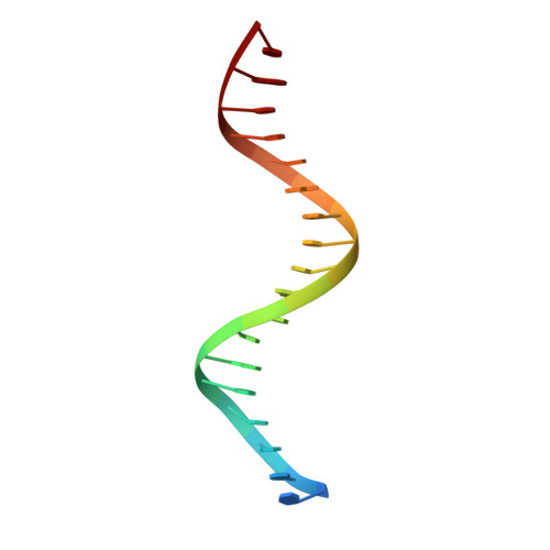
DNA (5'-D(P*CP*TP*CP*CP*TP*AP*TP*GP*AP*CP*TP*CP*GP*TP*CP*CP*AP*T)-3')A [auth C]19synthetic construct
Sequence Annotations
Expand
  • Reference Sequence

Find similar nucleic acids by:  Sequence   |   3D Structure  

Entity ID: 2
MoleculeChains LengthOrganismImage
DNA (5'-D(*AP*AP*TP*GP*GP*AP*(5CM)P*GP*AP*GP*TP*CP*AP*TP*AP*GP*GP*AP*G)-3')B [auth D]19synthetic construct
Sequence Annotations
Expand
  • Reference Sequence
Experimental Data & Validation

Experimental Data

  • Method: X-RAY DIFFRACTION
  • Resolution: 1.89 Å
  • R-Value Free: 0.227 
  • R-Value Work: 0.193 
  • R-Value Observed: 0.195 
  • Space Group: C 1 2 1
Unit Cell:
Length ( Å )Angle ( ˚ )
a = 158.871α = 90
b = 42.49β = 98.01
c = 45.167γ = 90
Software Package:
Software NamePurpose
PHENIXrefinement
PHENIXmodel building
SERGUIdata collection
HKL-2000data scaling
HKL-2000data reduction
PHENIXphasing

Structure Validation

View Full Validation Report


Entry History & Funding Information

Deposition Data


Funding OrganizationLocationGrant Number
National Institutes of Health/National Institute of General Medical Sciences (NIH/NIGMS)United States--

Revision History  (Full details and data files)

  • Version 1.0: 2017-03-01
    Type: Initial release
  • Version 1.1: 2017-05-03
    Changes: Database references
  • Version 1.2: 2017-09-27
    Changes: Data collection
  • Version 1.3: 2019-12-04
    Changes: Author supporting evidence
  • Version 1.4: 2023-10-04
    Changes: Data collection, Database references, Refinement description