5SZS

Glycan shield and epitope masking of a coronavirus spike protein observed by cryo-electron microscopy


Experimental Data Snapshot

  • Method: ELECTRON MICROSCOPY
  • Resolution: 3.40 Å
  • Aggregation State: PARTICLE 
  • Reconstruction Method: SINGLE PARTICLE 

wwPDB Validation   3D Report Full Report


This is version 2.0 of the entry. See complete history


Literature

Glycan shield and epitope masking of a coronavirus spike protein observed by cryo-electron microscopy.

Walls, A.C.Tortorici, M.A.Frenz, B.Snijder, J.Li, W.Rey, F.A.DiMaio, F.Bosch, B.J.Veesler, D.

(2016) Nat Struct Mol Biol 23: 899-905

  • DOI: https://doi.org/10.1038/nsmb.3293
  • Primary Citation of Related Structures:  
    5SZS

  • PubMed Abstract: 

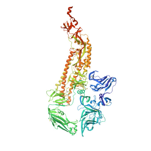
    The threat of a major coronavirus pandemic urges the development of strategies to combat these pathogens. Human coronavirus NL63 (HCoV-NL63) is an α-coronavirus that can cause severe lower-respiratory-tract infections requiring hospitalization. We report here the 3.4-Å-resolution cryo-EM reconstruction of the HCoV-NL63 coronavirus spike glycoprotein trimer, which mediates entry into host cells and is the main target of neutralizing antibodies during infection. The map resolves the extensive glycan shield obstructing the protein surface and, in combination with mass spectrometry, provides a structural framework to understand the accessibility to antibodies. The structure reveals the complete architecture of the fusion machinery including the triggering loop and the C-terminal domains, which contribute to anchoring the trimer to the viral membrane. Our data further suggest that HCoV-NL63 and other coronaviruses use molecular trickery, based on epitope masking with glycans and activating conformational changes, to evade the immune system of infected hosts.


  • Organizational Affiliation

    Department of Biochemistry, University of Washington, Seattle, Washington, USA.


Macromolecules
Find similar proteins by:  (by identity cutoff)  |  3D Structure
Entity ID: 1
MoleculeChains Sequence LengthOrganismDetailsImage
Spike glycoprotein
A, B, C
1,325Human coronavirus NL63Mutation(s): 0 
Gene Names: S2
UniProt
Find proteins for Q6Q1S2 (Human coronavirus NL63)
Explore Q6Q1S2 
Go to UniProtKB:  Q6Q1S2
Entity Groups  
Sequence Clusters30% Identity50% Identity70% Identity90% Identity95% Identity100% Identity
UniProt GroupQ6Q1S2
Sequence Annotations
Expand
  • Reference Sequence
Oligosaccharides

Help

Entity ID: 2
MoleculeChains Length2D Diagram Glycosylation3D Interactions
alpha-D-mannopyranose-(1-2)-alpha-D-mannopyranose-(1-3)-[alpha-D-mannopyranose-(1-6)]beta-D-mannopyranose-(1-4)-2-acetamido-2-deoxy-beta-D-glucopyranose-(1-4)-2-acetamido-2-deoxy-beta-D-glucopyranoseD,
TA [auth t],
Y
6N-Glycosylation
Glycosylation Resources
GlyTouCan:  G56014GC
GlyCosmos:  G56014GC
GlyGen:  G56014GC
Entity ID: 3
MoleculeChains Length2D Diagram Glycosylation3D Interactions
2-acetamido-2-deoxy-beta-D-glucopyranose-(1-4)-2-acetamido-2-deoxy-beta-D-glucopyranose
BA [auth b],
CB [auth 2],
DA [auth d],
E,
EA [auth e],
BA [auth b],
CB [auth 2],
DA [auth d],
E,
EA [auth e],
FB [auth 5],
G,
GB [auth 6],
HA [auth h],
HB [auth 7],
I,
IB [auth 8],
J,
JB [auth 9],
KA [auth k],
LA [auth l],
M,
MA [auth m],
MB [auth CA],
NA [auth n],
OA [auth o],
P,
Q,
R,
RA [auth r],
S,
T,
UA [auth u],
W,
WA [auth w],
YA [auth y],
Z,
ZA [auth z]
2N-Glycosylation
Glycosylation Resources
GlyTouCan:  G42666HT
GlyCosmos:  G42666HT
GlyGen:  G42666HT
Entity ID: 4
MoleculeChains Length2D Diagram Glycosylation3D Interactions
alpha-D-mannopyranose-(1-3)-beta-D-mannopyranose-(1-4)-2-acetamido-2-deoxy-beta-D-glucopyranose-(1-4)-2-acetamido-2-deoxy-beta-D-glucopyranose
AA [auth a],
F,
KB [auth AA],
PA [auth p],
U,
AA [auth a],
F,
KB [auth AA],
PA [auth p],
U,
VA [auth v]
4N-Glycosylation
Glycosylation Resources
GlyTouCan:  G81315DD
GlyCosmos:  G81315DD
GlyGen:  G81315DD
Entity ID: 5
MoleculeChains Length2D Diagram Glycosylation3D Interactions
alpha-D-mannopyranose-(1-6)-beta-D-mannopyranose-(1-4)-2-acetamido-2-deoxy-beta-D-glucopyranose-(1-4)-2-acetamido-2-deoxy-beta-D-glucopyranoseCA [auth c],
H,
XA [auth x]
4N-Glycosylation
Glycosylation Resources
GlyTouCan:  G22573RC
GlyCosmos:  G22573RC
GlyGen:  G22573RC
Entity ID: 6
MoleculeChains Length2D Diagram Glycosylation3D Interactions
alpha-D-mannopyranose-(1-2)-alpha-D-mannopyranose-(1-2)-alpha-D-mannopyranose-(1-3)-[alpha-D-mannopyranose-(1-3)-alpha-D-mannopyranose-(1-6)]beta-D-mannopyranose-(1-4)-2-acetamido-2-deoxy-beta-D-glucopyranose-(1-4)-2-acetamido-2-deoxy-beta-D-glucopyranoseAB [auth 0],
FA [auth f],
K
8N-Glycosylation
Glycosylation Resources
GlyTouCan:  G83582BK
GlyCosmos:  G83582BK
GlyGen:  G83582BK
Entity ID: 7
MoleculeChains Length2D Diagram Glycosylation3D Interactions
beta-D-mannopyranose-(1-4)-2-acetamido-2-deoxy-beta-D-glucopyranose-(1-4)-2-acetamido-2-deoxy-beta-D-glucopyranose
BB [auth 1],
EB [auth 4],
GA [auth g],
JA [auth j],
L,
BB [auth 1],
EB [auth 4],
GA [auth g],
JA [auth j],
L,
NB [auth DA],
O,
SA [auth s],
X
3N-Glycosylation
Glycosylation Resources
GlyTouCan:  G15407YE
GlyCosmos:  G15407YE
GlyGen:  G15407YE
Entity ID: 8
MoleculeChains Length2D Diagram Glycosylation3D Interactions
alpha-D-mannopyranose-(1-3)-[alpha-D-mannopyranose-(1-6)]beta-D-mannopyranose-(1-4)-2-acetamido-2-deoxy-beta-D-glucopyranose-(1-4)-2-acetamido-2-deoxy-beta-D-glucopyranose
DB [auth 3],
IA [auth i],
LB [auth BA],
N,
QA [auth q],
DB [auth 3],
IA [auth i],
LB [auth BA],
N,
QA [auth q],
V
5N-Glycosylation
Glycosylation Resources
GlyTouCan:  G22768VO
GlyCosmos:  G22768VO
GlyGen:  G22768VO
Small Molecules
Ligands 1 Unique
IDChains Name / Formula / InChI Key2D Diagram3D Interactions
NAG
Query on NAG

Download Ideal Coordinates CCD File 
AC [auth B]
BC [auth B]
CC [auth C]
DC [auth C]
EC [auth C]
AC [auth B],
BC [auth B],
CC [auth C],
DC [auth C],
EC [auth C],
FC [auth C],
GC [auth C],
HC [auth C],
IC [auth C],
OB [auth A],
PB [auth A],
QB [auth A],
RB [auth A],
SB [auth A],
TB [auth A],
UB [auth A],
VB [auth B],
WB [auth B],
XB [auth B],
YB [auth B],
ZB [auth B]
2-acetamido-2-deoxy-beta-D-glucopyranose
C8 H15 N O6
OVRNDRQMDRJTHS-FMDGEEDCSA-N
Experimental Data & Validation

Experimental Data

  • Method: ELECTRON MICROSCOPY
  • Resolution: 3.40 Å
  • Aggregation State: PARTICLE 
  • Reconstruction Method: SINGLE PARTICLE 
EM Software:
TaskSoftware PackageVersion
MODEL REFINEMENTRosetta
RECONSTRUCTIONRELION

Structure Validation

View Full Validation Report


Entry History & Funding Information

Deposition Data


Funding OrganizationLocationGrant Number
National Institutes of Health/National Institute of General Medical Sciences (NIH/NIGMS)United States1R01GM120553-01

Revision History  (Full details and data files)

  • Version 1.0: 2016-09-14
    Type: Initial release
  • Version 1.1: 2016-09-21
    Changes: Database references
  • Version 1.2: 2016-09-28
    Changes: Database references
  • Version 1.3: 2016-10-19
    Changes: Database references
  • Version 1.4: 2017-09-20
    Changes: Author supporting evidence, Data collection
  • Version 1.5: 2017-11-08
    Changes: Derived calculations
  • Version 1.6: 2019-12-25
    Changes: Author supporting evidence
  • Version 2.0: 2020-07-29
    Type: Remediation
    Reason: Carbohydrate remediation
    Changes: Atomic model, Data collection, Derived calculations, Other, Structure summary