5SYN

Cocrystal structure of the human acyl protein thioesterase 2 with an isoform-selective inhibitor, ML349


Experimental Data Snapshot

  • Method: X-RAY DIFFRACTION
  • Resolution: 1.64 Å
  • R-Value Free: 0.250 
  • R-Value Work: 0.220 

wwPDB Validation   3D Report Full Report


Ligand Structure Quality Assessment 


This is version 1.5 of the entry. See complete history


Literature

Molecular Mechanism for Isoform-Selective Inhibition of Acyl Protein Thioesterases 1 and 2 (APT1 and APT2).

Won, S.J.Davda, D.Labby, K.J.Hwang, S.Y.Pricer, R.Majmudar, J.D.Armacost, K.A.Rodriguez, L.A.Rodriguez, C.L.Chong, F.S.Torossian, K.A.Palakurthi, J.Hur, E.S.Meagher, J.L.Brooks, C.L.Stuckey, J.A.Martin, B.R.

(2016) ACS Chem Biol 11: 3374-3382

  • DOI: https://doi.org/10.1021/acschembio.6b00720
  • Primary Citation of Related Structures:  
    5SYM, 5SYN

  • PubMed Abstract: 

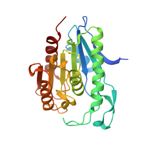
    Post-translational S-palmitoylation directs the trafficking and membrane localization of hundreds of cellular proteins, often involving a coordinated palmitoylation cycle that requires both protein acyl transferases (PATs) and acyl protein thioesterases (APTs) to actively redistribute S-palmitoylated proteins toward different cellular membrane compartments. This process is necessary for the trafficking and oncogenic signaling of S-palmitoylated Ras isoforms, and potentially many peripheral membrane proteins. The depalmitoylating enzymes APT1 and APT2 are separately conserved in all vertebrates, suggesting unique functional roles for each enzyme. The recent discovery of the APT isoform-selective inhibitors ML348 and ML349 has opened new possibilities to probe the function of each enzyme, yet it remains unclear how each inhibitor achieves orthogonal inhibition. Herein, we report the high-resolution structure of human APT2 in complex with ML349 (1.64 Å), as well as the complementary structure of human APT1 bound to ML348 (1.55 Å). Although the overall peptide backbone structures are nearly identical, each inhibitor adopts a distinct conformation within each active site. In APT1, the trifluoromethyl group of ML348 is positioned above the catalytic triad, but in APT2, the sulfonyl group of ML349 forms hydrogen bonds with active site resident waters to indirectly engage the catalytic triad and oxyanion hole. Reciprocal mutagenesis and activity profiling revealed several differing residues surrounding the active site that serve as critical gatekeepers for isoform accessibility and dynamics. Structural and biochemical analysis suggests the inhibitors occupy a putative acyl-binding region, establishing the mechanism for isoform-specific inhibition, hydrolysis of acyl substrates, and structural orthogonality important for future probe development.


  • Organizational Affiliation

    Program in Chemical Biology, ‡Department of Chemistry, §Department of Biophysics, and ∥Life Sciences Institute, University of Michigan , 930 N. University Ave., Ann Arbor, Michigan 48109, United States.


Macromolecules
Find similar proteins by:  (by identity cutoff)  |  3D Structure
Entity ID: 1
MoleculeChains Sequence LengthOrganismDetailsImage
Acyl-protein thioesterase 2
A, B, C, D
231Homo sapiensMutation(s): 0 
Gene Names: LYPLA2APT2
EC: 3.1.2
UniProt & NIH Common Fund Data Resources
Find proteins for O95372 (Homo sapiens)
Explore O95372 
Go to UniProtKB:  O95372
GTEx:  ENSG00000011009 
Entity Groups  
Sequence Clusters30% Identity50% Identity70% Identity90% Identity95% Identity100% Identity
UniProt GroupO95372
Sequence Annotations
Expand
  • Reference Sequence
Small Molecules
Ligands 2 Unique
IDChains Name / Formula / InChI Key2D Diagram3D Interactions
71T
Query on 71T

Download Ideal Coordinates CCD File 
E [auth A],
G [auth B],
L [auth C],
N [auth D]
2-[4-(4-methoxyphenyl)piperazine-1-carbonyl]-5lambda~6~-thieno[3,2-c][1]benzothiopyran-5,5(4H)-dione
C23 H22 N2 O4 S2
YVIJPELUPZUEJX-UHFFFAOYSA-N
EDO
Query on EDO

Download Ideal Coordinates CCD File 
F [auth A]
H [auth B]
I [auth B]
J [auth B]
K [auth B]
F [auth A],
H [auth B],
I [auth B],
J [auth B],
K [auth B],
M [auth C]
1,2-ETHANEDIOL
C2 H6 O2
LYCAIKOWRPUZTN-UHFFFAOYSA-N
Binding Affinity Annotations 
IDSourceBinding Affinity
71T BindingDB:  5SYN Ki: min: 120, max: 1.00e+4 (nM) from 8 assay(s)
Kd: min: 160, max: 240 (nM) from 2 assay(s)
IC50: min: 1000, max: 1100 (nM) from 2 assay(s)
Binding MOAD:  5SYN Kd: 240 (nM) from 1 assay(s)
Experimental Data & Validation

Experimental Data

  • Method: X-RAY DIFFRACTION
  • Resolution: 1.64 Å
  • R-Value Free: 0.250 
  • R-Value Work: 0.220 
  • Space Group: C 1 2 1
Unit Cell:
Length ( Å )Angle ( ˚ )
a = 78.21α = 90
b = 79.79β = 93.3
c = 138.61γ = 90
Software Package:
Software NamePurpose
Cootmodel building
BUSTERrefinement
SCALAdata scaling
BALBESphasing
MOSFLMdata reduction

Structure Validation

View Full Validation Report


Ligand Structure Quality Assessment 


Entry History & Funding Information

Deposition Data


Funding OrganizationLocationGrant Number
National Institutes of Health/National Cancer Institute (NIH/NCI)United StatesR00 CA151460
National Institutes of Health/National Institute of General Medical Sciences (NIH/NIGMS)United StatesDP2 GM114848

Revision History  (Full details and data files)

  • Version 1.0: 2016-10-26
    Type: Initial release
  • Version 1.1: 2016-11-09
    Changes: Database references
  • Version 1.2: 2016-12-28
    Changes: Database references
  • Version 1.3: 2017-09-20
    Changes: Author supporting evidence, Refinement description
  • Version 1.4: 2019-12-04
    Changes: Author supporting evidence
  • Version 1.5: 2023-10-04
    Changes: Advisory, Data collection, Database references, Refinement description