5SV6

Crystal structure of MxaJ from Methlophaga aminisulfidivorans MPT


Experimental Data Snapshot

  • Method: X-RAY DIFFRACTION
  • Resolution: 1.92 Å
  • R-Value Free: 0.239 
  • R-Value Work: 0.178 
  • R-Value Observed: 0.184 

wwPDB Validation   3D Report Full Report


This is version 1.0 of the entry. See complete history


Literature

MxaJ structure reveals a periplasmic binding protein-like architecture with unique secondary structural elements

Choi, J.M.Cao, T.P.Kim, S.W.Lee, K.H.Lee, S.H.

(2017) Proteins 85: 1379-1386

  • DOI: https://doi.org/10.1002/prot.25283
  • Primary Citation of Related Structures:  
    5SV6

  • PubMed Abstract: 

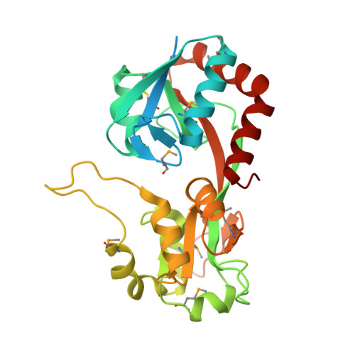
    MxaJ is a component of type II methanol dehydrogenase (MDH) that mediates electron transfer during methanol oxidation in methanotrophic bacteria. However, little is known about how MxaJ structurally cooperates with MDH and Cytochrome c L . Here, we report for the first time the crystal structure of MxaJ. MxaJ consists of eight α-helices and six β-strands, and resembles the "bi-lobate" folding architecture found in periplasmic binding proteins. Distinctive features of MxaJ include prominent loops and a β-strand around the hinge region supporting the ligand-binding cavity, which might provide a more favorable framework for interacting with proteins rather than small molecules. Proteins 2017; 85:1379-1386. © 2017 Wiley Periodicals, Inc.


  • Organizational Affiliation

    Department of Cellular and Molecular Medicine, Chosun University School of Medicine, Gwangju, 61452, Korea.


Macromolecules
Find similar proteins by:  (by identity cutoff)  |  3D Structure
Entity ID: 1
MoleculeChains Sequence LengthOrganismDetailsImage
Extracellular solute-binding protein, family 3291Methylophaga aminisulfidivorans MPMutation(s): 0 
Gene Names: mxaJ
UniProt
Find proteins for A3FJ49 (Methylophaga aminisulfidivorans MP)
Explore A3FJ49 
Go to UniProtKB:  A3FJ49
Entity Groups  
Sequence Clusters30% Identity50% Identity70% Identity90% Identity95% Identity100% Identity
UniProt GroupA3FJ49
Sequence Annotations
Expand
  • Reference Sequence
Small Molecules
Ligands 1 Unique
IDChains Name / Formula / InChI Key2D Diagram3D Interactions
BR
Query on BR

Download Ideal Coordinates CCD File 
B [auth A]BROMIDE ION
Br
CPELXLSAUQHCOX-UHFFFAOYSA-M
Modified Residues  1 Unique
IDChains TypeFormula2D DiagramParent
MSE
Query on MSE
A
L-PEPTIDE LINKINGC5 H11 N O2 SeMET
Experimental Data & Validation

Experimental Data

  • Method: X-RAY DIFFRACTION
  • Resolution: 1.92 Å
  • R-Value Free: 0.239 
  • R-Value Work: 0.178 
  • R-Value Observed: 0.184 
  • Space Group: P 21 21 21
Unit Cell:
Length ( Å )Angle ( ˚ )
a = 37.127α = 90
b = 63.761β = 90
c = 99.246γ = 90
Software Package:
Software NamePurpose
PHENIXrefinement
HKL-2000data reduction
HKL-2000data scaling
PHENIXphasing

Structure Validation

View Full Validation Report


Entry History 

Deposition Data

Revision History  (Full details and data files)

  • Version 1.0: 2017-07-12
    Type: Initial release