


Crystal structure of human Cathepsin-S with bound ligand
Bembenek, S.D., Ameriks, M.K., Mirzadegan, T., Yang, H., Shao, C., Burley, S.K.To be published.
Experimental Data Snapshot
Entity ID: 1 | |||||
|---|---|---|---|---|---|
| Molecule | Chains | Sequence Length | Organism | Details | Image |
| Cathepsin S | 223 | Homo sapiens | Mutation(s): 1 Gene Names: CTSS EC: 3.4.22.27 | 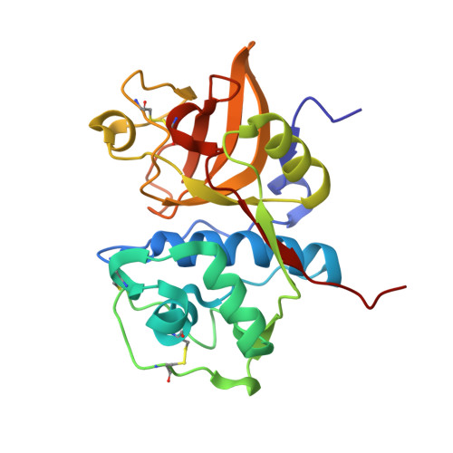 | |
UniProt & NIH Common Fund Data Resources | |||||
Find proteins for P25774 (Homo sapiens) Explore P25774 Go to UniProtKB: P25774 | |||||
PHAROS: P25774 GTEx: ENSG00000163131 | |||||
Entity Groups | |||||
| Sequence Clusters | 30% Identity50% Identity70% Identity90% Identity95% Identity100% Identity | ||||
| UniProt Group | P25774 | ||||
Sequence AnnotationsExpand | |||||
| |||||
| Ligands 3 Unique | |||||
|---|---|---|---|---|---|
| ID | Chains | Name / Formula / InChI Key | 2D Diagram | 3D Interactions | |
| BJS Query on BJS | F [auth A], I [auth B], K [auth C], N [auth D] | 2-(3-[3-({3-[(benzylamino)methyl]-4-chlorophenyl}ethynyl)-4-chlorophenyl]-1-{3-[(3S)-3-methylmorpholin-4-yl]propyl}-1,4,6,7-tetrahydro-5H-pyrazolo[4,3-c]pyridin-5-yl)-2-oxoacetamide C38 H40 Cl2 N6 O3 CIONECARUUEYBQ-SANMLTNESA-N |  | ||
| SO4 Query on SO4 | E [auth A], G [auth B], J [auth C], L [auth D] | SULFATE ION O4 S QAOWNCQODCNURD-UHFFFAOYSA-L |  | ||
| GOL Query on GOL | H [auth B], M [auth D] | GLYCEROL C3 H8 O3 PEDCQBHIVMGVHV-UHFFFAOYSA-N |  | ||
| Length ( Å ) | Angle ( ˚ ) |
|---|---|
| a = 54.12 | α = 88.06 |
| b = 68.13 | β = 87.69 |
| c = 79.02 | γ = 73.08 |
| Software Name | Purpose |
|---|---|
| BUSTER | refinement |
| PDB_EXTRACT | data extraction |
| MOSFLM | data reduction |
| SCALA | data scaling |
RCSB PDB (citation) is hosted by
RCSB PDB is a member of the

