


Crystal Structure of a Factor VIIa complex
Mayweg, A., Roever, S., Rudolph, M.G.To be published.
Experimental Data Snapshot
Entity ID: 1 | |||||
|---|---|---|---|---|---|
| Molecule | Chains | Sequence Length | Organism | Details | Image |
| Coagulation factor VII heavy chain | A [auth H] | 254 | Homo sapiens | Mutation(s): 0 Gene Names: F7 EC: 3.4.21.21 | 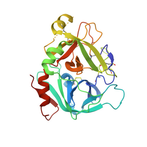 |
UniProt & NIH Common Fund Data Resources | |||||
Find proteins for P08709 (Homo sapiens) Explore P08709 Go to UniProtKB: P08709 | |||||
PHAROS: P08709 GTEx: ENSG00000057593 | |||||
Entity Groups | |||||
| Sequence Clusters | 30% Identity50% Identity70% Identity90% Identity95% Identity100% Identity | ||||
| UniProt Group | P08709 | ||||
Sequence AnnotationsExpand | |||||
| |||||
Entity ID: 2 | |||||
|---|---|---|---|---|---|
| Molecule | Chains | Sequence Length | Organism | Details | Image |
| Coagulation factor VII light chain | B [auth L] | 64 | Homo sapiens | Mutation(s): 0 Gene Names: F7 EC: 3.4.21.21 | 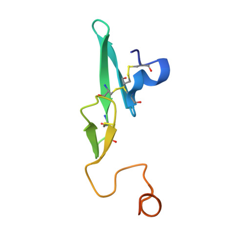 |
UniProt & NIH Common Fund Data Resources | |||||
Find proteins for P08709 (Homo sapiens) Explore P08709 Go to UniProtKB: P08709 | |||||
PHAROS: P08709 GTEx: ENSG00000057593 | |||||
Entity Groups | |||||
| Sequence Clusters | 30% Identity50% Identity70% Identity90% Identity95% Identity100% Identity | ||||
| UniProt Group | P08709 | ||||
Sequence AnnotationsExpand | |||||
| |||||
| Ligands 5 Unique | |||||
|---|---|---|---|---|---|
| ID | Chains | Name / Formula / InChI Key | 2D Diagram | 3D Interactions | |
| 7XD Query on 7XD | C [auth H] | 1-[[3-[2-oxidanyl-3-(1~{H}-pyrrolo[3,2-c]pyridin-2-yl)phenyl]phenyl]methyl]-3-phenyl-urea C27 H22 N4 O2 MYWMHTLQFNYXPC-UHFFFAOYSA-N |  | ||
| SO4 Query on SO4 | K [auth H], M [auth L] | SULFATE ION O4 S QAOWNCQODCNURD-UHFFFAOYSA-L |  | ||
| GOL Query on GOL | E [auth H], F [auth H], G [auth H], L | GLYCEROL C3 H8 O3 PEDCQBHIVMGVHV-UHFFFAOYSA-N |  | ||
| CA Query on CA | H | CALCIUM ION Ca BHPQYMZQTOCNFJ-UHFFFAOYSA-N |  | ||
| CL Query on CL | D [auth H], I [auth H], J [auth H] | CHLORIDE ION Cl VEXZGXHMUGYJMC-UHFFFAOYSA-M |  | ||
| Modified Residues 1 Unique | |||||
|---|---|---|---|---|---|
| ID | Chains | Type | Formula | 2D Diagram | Parent |
| MHO Query on MHO | A [auth H] | L-PEPTIDE LINKING | C5 H11 N O3 S |  | MET |
| Length ( Å ) | Angle ( ˚ ) |
|---|---|
| a = 95.217 | α = 90 |
| b = 95.217 | β = 90 |
| c = 116.197 | γ = 90 |
| Software Name | Purpose |
|---|---|
| SCALEPACK | data scaling |
| REFMAC | refinement |
| PDB_EXTRACT | data extraction |
| XDS | data reduction |
| PHASER | phasing |
RCSB PDB (citation) is hosted by
RCSB PDB is a member of the

