5OYO

Crystal structure of BlaC from Mycobacterium tuberculosis


Experimental Data Snapshot

  • Method: X-RAY DIFFRACTION
  • Resolution: 2.10 Å
  • R-Value Free: 0.250 
  • R-Value Work: 0.206 
  • R-Value Observed: 0.208 

wwPDB Validation   3D Report Full Report


This is version 1.2 of the entry. See complete history


Literature

Phosphate Promotes the Recovery of Mycobacterium tuberculosis beta-Lactamase from Clavulanic Acid Inhibition.

Elings, W.Tassoni, R.van der Schoot, S.A.Luu, W.Kynast, J.P.Dai, L.Blok, A.J.Timmer, M.Florea, B.I.Pannu, N.S.Ubbink, M.

(2017) Biochemistry 56: 6257-6267

  • DOI: https://doi.org/10.1021/acs.biochem.7b00556
  • Primary Citation of Related Structures:  
    5NJ2, 5OYO

  • PubMed Abstract: 

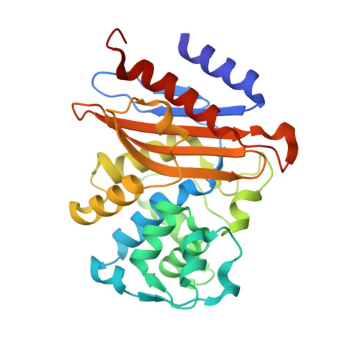
    The rise of multi- and even totally antibiotic resistant forms of Mycobacterium tuberculosis underlines the need for new antibiotics. The pathogen is resistant to β-lactam compounds due to its native serine β-lactamase, BlaC. This resistance can be circumvented by administration of a β-lactamase inhibitor. We studied the interaction between BlaC and the inhibitor clavulanic acid. Our data show hydrolysis of clavulanic acid and recovery of BlaC activity upon prolonged incubation. The rate of clavulanic acid hydrolysis is much higher in the presence of phosphate ions. A specific binding site for phosphate is identified in the active site pocket, both in the crystalline state and in solution. NMR spectroscopy experiments show that phosphate binds to this site with a dissociation constant of 30 mM in the free enzyme. We conclude that inhibition of BlaC by clavulanic acid is reversible and that phosphate ions can promote the hydrolysis of the inhibitor.


  • Organizational Affiliation

    Leiden Institute of Chemistry, Leiden University , Einsteinweg 55, Leiden, The Netherlands.


Macromolecules
Find similar proteins by:  (by identity cutoff)  |  3D Structure
Entity ID: 1
MoleculeChains Sequence LengthOrganismDetailsImage
Beta-lactamase
A, B
274Mycobacterium tuberculosisMutation(s): 0 
Gene Names: blaCERS027646_02769
EC: 3.5.2.6
UniProt
Find proteins for P9WKD3 (Mycobacterium tuberculosis (strain ATCC 25618 / H37Rv))
Explore P9WKD3 
Go to UniProtKB:  P9WKD3
Entity Groups  
Sequence Clusters30% Identity50% Identity70% Identity90% Identity95% Identity100% Identity
UniProt GroupP9WKD3
Sequence Annotations
Expand
  • Reference Sequence
Small Molecules
Ligands 3 Unique
IDChains Name / Formula / InChI Key2D Diagram3D Interactions
PGE
Query on PGE

Download Ideal Coordinates CCD File 
H [auth A],
L [auth B],
M [auth B]
TRIETHYLENE GLYCOL
C6 H14 O4
ZIBGPFATKBEMQZ-UHFFFAOYSA-N
GOL
Query on GOL

Download Ideal Coordinates CCD File 
G [auth A]GLYCEROL
C3 H8 O3
PEDCQBHIVMGVHV-UHFFFAOYSA-N
ACT
Query on ACT

Download Ideal Coordinates CCD File 
C [auth A]
D [auth A]
E [auth A]
F [auth A]
I [auth B]
C [auth A],
D [auth A],
E [auth A],
F [auth A],
I [auth B],
J [auth B],
K [auth B]
ACETATE ION
C2 H3 O2
QTBSBXVTEAMEQO-UHFFFAOYSA-M
Experimental Data & Validation

Experimental Data

  • Method: X-RAY DIFFRACTION
  • Resolution: 2.10 Å
  • R-Value Free: 0.250 
  • R-Value Work: 0.206 
  • R-Value Observed: 0.208 
  • Space Group: P 1
Unit Cell:
Length ( Å )Angle ( ˚ )
a = 39.589α = 104.29
b = 41.417β = 90.07
c = 76.523γ = 90.67
Software Package:
Software NamePurpose
REFMACrefinement
MOLREPphasing

Structure Validation

View Full Validation Report


Entry History 

Deposition Data

Revision History  (Full details and data files)

  • Version 1.0: 2017-11-15
    Type: Initial release
  • Version 1.1: 2017-12-06
    Changes: Database references
  • Version 1.2: 2024-01-17
    Changes: Data collection, Database references, Refinement description