Activation mechanism of the calcium-activated chloride channel TMEM16A revealed by cryo-EM.
Paulino, C., Kalienkova, V., Lam, A.K.M., Neldner, Y., Dutzler, R.(2017) Nature 552: 421-425
- PubMed: 29236691
- DOI: https://doi.org/10.1038/nature24652
- Primary Citation of Related Structures:
5OYB, 5OYG - PubMed Abstract:
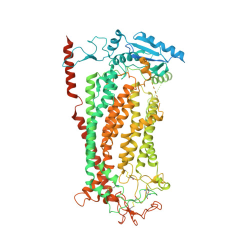
The calcium-activated chloride channel TMEM16A is a ligand-gated anion channel that opens in response to an increase in intracellular Ca 2+ concentration. The protein is broadly expressed and contributes to diverse physiological processes, including transepithelial chloride transport and the control of electrical signalling in smooth muscles and certain neurons. As a member of the TMEM16 (or anoctamin) family of membrane proteins, TMEM16A is closely related to paralogues that function as scramblases, which facilitate the bidirectional movement of lipids across membranes. The unusual functional diversity of the TMEM16 family and the relationship between two seemingly incompatible transport mechanisms has been the focus of recent investigations. Previous breakthroughs were obtained from the X-ray structure of the lipid scramblase of the fungus Nectria haematococca (nhTMEM16), and from the cryo-electron microscopy structure of mouse TMEM16A at 6.6 Å (ref. 14). Although the latter structure disclosed the architectural differences that distinguish ion channels from lipid scramblases, its low resolution did not permit a detailed molecular description of the protein or provide any insight into its activation by Ca 2+ . Here we describe the structures of mouse TMEM16A at high resolution in the presence and absence of Ca 2+ . These structures reveal the differences between ligand-bound and ligand-free states of a calcium-activated chloride channel, and when combined with functional experiments suggest a mechanism for gating. During activation, the binding of Ca 2+ to a site located within the transmembrane domain, in the vicinity of the pore, alters the electrostatic properties of the ion conduction path and triggers a conformational rearrangement of an α-helix that comes into physical contact with the bound ligand, and thereby directly couples ligand binding and pore opening. Our study describes a process that is unique among channel proteins, but one that is presumably general for both functional branches of the TMEM16 family.
Organizational Affiliation:
Department of Biochemistry, University of Zurich, Winterthurerstrasse 190, CH-8057 Zurich, Switzerland.