5OX6

HIF prolyl hydroxylase 2 (PHD2/ EGLN1) in complex with Vadadustat


Experimental Data Snapshot

  • Method: X-RAY DIFFRACTION
  • Resolution: 1.99 Å
  • R-Value Free: 0.205 
  • R-Value Work: 0.174 
  • R-Value Observed: 0.176 

wwPDB Validation   3D Report Full Report


Ligand Structure Quality Assessment 


This is version 1.2 of the entry. See complete history


Literature

Molecular and cellular mechanisms of HIF prolyl hydroxylase inhibitors in clinical trials.

Yeh, T.L.Leissing, T.M.Abboud, M.I.Thinnes, C.C.Atasoylu, O.Holt-Martyn, J.P.Zhang, D.Tumber, A.Lippl, K.Lohans, C.T.Leung, I.K.H.Morcrette, H.Clifton, I.J.Claridge, T.D.W.Kawamura, A.Flashman, E.Lu, X.Ratcliffe, P.J.Chowdhury, R.Pugh, C.W.Schofield, C.J.

(2017) Chem Sci 8: 7651-7668

  • DOI: https://doi.org/10.1039/c7sc02103h
  • Primary Citation of Related Structures:  
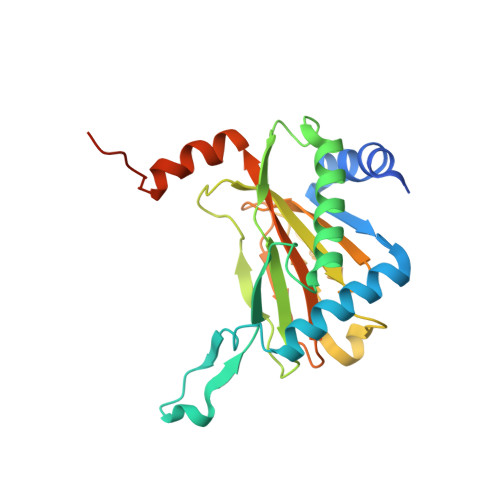
    5OP6, 5OP8, 5OPC, 5OX5, 5OX6

  • PubMed Abstract: 

    Inhibition of the human 2-oxoglutarate (2OG) dependent hypoxia inducible factor (HIF) prolyl hydroxylases (human PHD1-3) causes upregulation of HIF, thus promoting erythropoiesis and is therefore of therapeutic interest. We describe cellular, biophysical, and biochemical studies comparing four PHD inhibitors currently in clinical trials for anaemia treatment, that describe their mechanisms of action, potency against isolated enzymes and in cells, and selectivities versus representatives of other human 2OG oxygenase subfamilies. The 'clinical' PHD inhibitors are potent inhibitors of PHD catalyzed hydroxylation of the HIF-α oxygen dependent degradation domains (ODDs), and selective against most, but not all, representatives of other human 2OG dependent dioxygenase subfamilies. Crystallographic and NMR studies provide insights into the different active site binding modes of the inhibitors. Cell-based results reveal the inhibitors have similar effects on the upregulation of HIF target genes, but differ in the kinetics of their effects and in extent of inhibition of hydroxylation of the N- and C-terminal ODDs; the latter differences correlate with the biophysical observations.


  • Organizational Affiliation

    Chemistry Research Laboratory , Department of Chemistry , University of Oxford , Oxford OX1 3TA , UK . Email: christopher.schofield@chem.ox.ac.uk.


Macromolecules
Find similar proteins by:  (by identity cutoff)  |  3D Structure
Entity ID: 1
MoleculeChains Sequence LengthOrganismDetailsImage
Egl nine homolog 1252Homo sapiensMutation(s): 0 
Gene Names: EGLN1C1orf12PNAS-118PNAS-137
EC: 1.14.11.29
UniProt & NIH Common Fund Data Resources
Find proteins for Q9GZT9 (Homo sapiens)
Explore Q9GZT9 
Go to UniProtKB:  Q9GZT9
PHAROS:  Q9GZT9
GTEx:  ENSG00000135766 
Entity Groups  
Sequence Clusters30% Identity50% Identity70% Identity90% Identity95% Identity100% Identity
UniProt GroupQ9GZT9
Sequence Annotations
Expand
  • Reference Sequence
Small Molecules
Binding Affinity Annotations 
IDSourceBinding Affinity
A1Z Binding MOAD:  5OX6 IC50: 29 (nM) from 1 assay(s)
BindingDB:  5OX6 IC50: min: 29, max: 1.37e+4 (nM) from 4 assay(s)
Experimental Data & Validation

Experimental Data

  • Method: X-RAY DIFFRACTION
  • Resolution: 1.99 Å
  • R-Value Free: 0.205 
  • R-Value Work: 0.174 
  • R-Value Observed: 0.176 
  • Space Group: P 63
Unit Cell:
Length ( Å )Angle ( ˚ )
a = 111.088α = 90
b = 111.088β = 90
c = 39.971γ = 120
Software Package:
Software NamePurpose
PHENIXrefinement
HKL-2000data reduction
HKL-2000data scaling
PHASERphasing

Structure Validation

View Full Validation Report


Ligand Structure Quality Assessment 


Entry History 

Deposition Data

Revision History  (Full details and data files)

  • Version 1.0: 2017-10-18
    Type: Initial release
  • Version 1.1: 2018-02-21
    Changes: Database references
  • Version 1.2: 2024-01-17
    Changes: Data collection, Database references, Derived calculations, Refinement description