5OVX

X-Ray Characterization of Striatal-Enriched Protein Tyrosine Phosphatase Inhibitors


Experimental Data Snapshot

  • Method: X-RAY DIFFRACTION
  • Resolution: 2.10 Å
  • R-Value Free: 0.228 
  • R-Value Work: 0.195 
  • R-Value Observed: 0.197 

wwPDB Validation   3D Report Full Report


Ligand Structure Quality Assessment 


This is version 1.2 of the entry. See complete history


Literature

X-ray Characterization and Structure-Based Optimization of Striatal-Enriched Protein Tyrosine Phosphatase Inhibitors.

Witten, M.R.Wissler, L.Snow, M.Geschwindner, S.Read, J.A.Brandon, N.J.Nairn, A.C.Lombroso, P.J.Kack, H.Ellman, J.A.

(2017) J Med Chem 60: 9299-9319

  • DOI: https://doi.org/10.1021/acs.jmedchem.7b01292
  • Primary Citation of Related Structures:  
    5OVR, 5OVX, 5OW1

  • PubMed Abstract: 

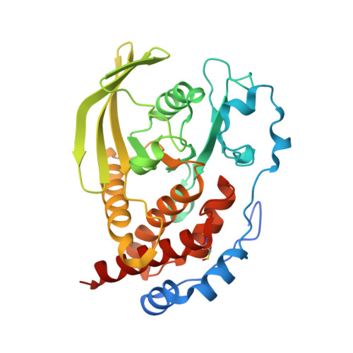
    Excessive activity of striatal-enriched protein tyrosine phosphatase (STEP) in the brain has been detected in numerous neuropsychiatric disorders including Alzheimer's disease. Notably, knockdown of STEP in an Alzheimer mouse model effected an increase in the phosphorylation levels of downstream STEP substrates and a significant reversal in the observed cognitive and memory deficits. These data point to the promising potential of STEP as a target for drug discovery in Alzheimer's treatment. We previously reported a substrate-based approach to the development of low molecular weight STEP inhibitors with K i values as low as 7.8 μM. Herein, we disclose the first X-ray crystal structures of inhibitors bound to STEP and the surprising finding that they occupy noncoincident binding sites. Moreover, we utilize this structural information to optimize the inhibitor structure to achieve a K i of 110 nM, with 15-60-fold selectivity across a series of phosphatases.


  • Organizational Affiliation

    Department of Chemistry, Yale University , New Haven, Connecticut 06520, United States.


Macromolecules
Find similar proteins by:  (by identity cutoff)  |  3D Structure
Entity ID: 1
MoleculeChains Sequence LengthOrganismDetailsImage
Tyrosine-protein phosphatase non-receptor type 5305Homo sapiensMutation(s): 0 
Gene Names: PTPN5
EC: 3.1.3.48
UniProt & NIH Common Fund Data Resources
Find proteins for P54829 (Homo sapiens)
Explore P54829 
Go to UniProtKB:  P54829
PHAROS:  P54829
GTEx:  ENSG00000110786 
Entity Groups  
Sequence Clusters30% Identity50% Identity70% Identity90% Identity95% Identity100% Identity
UniProt GroupP54829
Sequence Annotations
Expand
  • Reference Sequence
Small Molecules
Ligands 1 Unique
IDChains Name / Formula / InChI Key2D Diagram3D Interactions
AY5
Query on AY5

Download Ideal Coordinates CCD File 
B [auth A][(~{S})-[4-[3-[(~{S})-(3,4-dichlorophenyl)-oxidanyl-methyl]phenyl]phenyl]-oxidanyl-methyl]phosphonic acid
C20 H17 Cl2 O5 P
KYNFSOSPDJUFDE-PMACEKPBSA-N
Modified Residues  1 Unique
IDChains TypeFormula2D DiagramParent
CSO
Query on CSO
A
L-PEPTIDE LINKINGC3 H7 N O3 SCYS
Binding Affinity Annotations 
IDSourceBinding Affinity
AY5 Binding MOAD:  5OVX Kd: 3.14e+4 (nM) from 1 assay(s)
Experimental Data & Validation

Experimental Data

  • Method: X-RAY DIFFRACTION
  • Resolution: 2.10 Å
  • R-Value Free: 0.228 
  • R-Value Work: 0.195 
  • R-Value Observed: 0.197 
  • Space Group: P 21 21 21
Unit Cell:
Length ( Å )Angle ( ˚ )
a = 52.117α = 90
b = 64.124β = 90
c = 100.338γ = 90
Software Package:
Software NamePurpose
Aimlessdata scaling
BUSTERrefinement
PDB_EXTRACTdata extraction
XDSdata reduction
PHASERphasing

Structure Validation

View Full Validation Report


Ligand Structure Quality Assessment 


Entry History 

Deposition Data

Revision History  (Full details and data files)

  • Version 1.0: 2017-11-22
    Type: Initial release
  • Version 1.1: 2017-11-29
    Changes: Database references
  • Version 1.2: 2024-01-17
    Changes: Data collection, Database references, Refinement description