5OV3

Structure of the RbBP5 beta-propeller domain


Experimental Data Snapshot

  • Method: X-RAY DIFFRACTION
  • Resolution: 2.45 Å
  • R-Value Free: 0.271 
  • R-Value Work: 0.196 
  • R-Value Observed: 0.200 

wwPDB Validation   3D Report Full Report


This is version 1.3 of the entry. See complete history


Literature

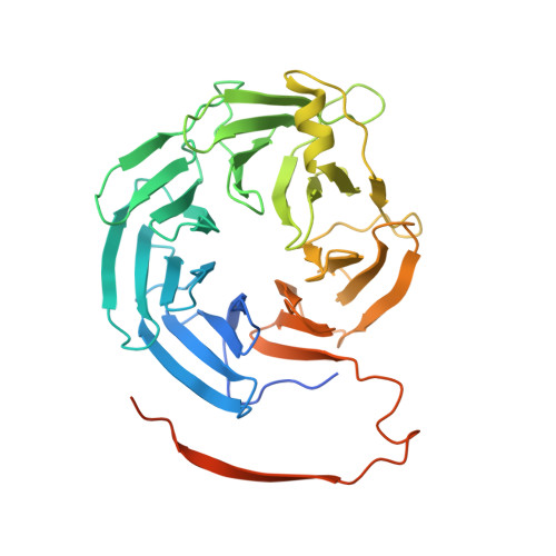
The structure of the RbBP5 beta-propeller domain reveals a surface with potential nucleic acid binding sites.

Mittal, A.Hobor, F.Zhang, Y.Martin, S.R.Gamblin, S.J.Ramos, A.Wilson, J.R.

(2018) Nucleic Acids Res 46: 3802-3812

  • DOI: https://doi.org/10.1093/nar/gky199
  • Primary Citation of Related Structures:  
    5OV3

  • PubMed Abstract: 

    The multi-protein complex WRAD, formed by WDR5, RbBP5, Ash2L and Dpy30, binds to the MLL SET domain to stabilize the catalytically active conformation required for histone H3K4 methylation. In addition, the WRAD complex contributes to the targeting of the activated complex to specific sites on chromatin. RbBP5 is central to MLL catalytic activation, by making critical contacts with the other members of the complex. Interestingly its only major structural domain, a canonical WD40 repeat β-propeller, is not implicated in this function. Here, we present the structure of the RbBP5 β-propeller domain revealing a distinct, feature rich surface, dominated by clusters of Arginine residues. Our nuclear magnetic resonance binding data supports the hypothesis that in addition to the role of RbBP5 in catalytic activation, its β-propeller domain is a platform for the recruitment of the MLL complexes to chromatin targets through its direct interaction with nucleic acids.


  • Organizational Affiliation

    The Francis Crick Institute, 1 Midland Road, London NW1 1AT, UK.


Macromolecules
Find similar proteins by:  (by identity cutoff)  |  3D Structure
Entity ID: 1
MoleculeChains Sequence LengthOrganismDetailsImage
Retinoblastoma-binding protein 5
A, B
381Mus musculusMutation(s): 1 
Gene Names: Rbbp5
UniProt
Find proteins for Q8BX09 (Mus musculus)
Explore Q8BX09 
Go to UniProtKB:  Q8BX09
Entity Groups  
Sequence Clusters30% Identity50% Identity70% Identity90% Identity95% Identity100% Identity
UniProt GroupQ8BX09
Sequence Annotations
Expand
  • Reference Sequence

Find similar proteins by:  Sequence   |   3D Structure  

Entity ID: 2
MoleculeChains Sequence LengthOrganismDetailsImage
Retinoblastoma-binding protein 56Mus musculusMutation(s): 1 
Gene Names: Rbbp5
UniProt
Find proteins for Q8BX09 (Mus musculus)
Explore Q8BX09 
Go to UniProtKB:  Q8BX09
Entity Groups  
UniProt GroupQ8BX09
Sequence Annotations
Expand
  • Reference Sequence
Small Molecules
Ligands 2 Unique
IDChains Name / Formula / InChI Key2D Diagram3D Interactions
PG4
Query on PG4

Download Ideal Coordinates CCD File 
E [auth B]TETRAETHYLENE GLYCOL
C8 H18 O5
UWHCKJMYHZGTIT-UHFFFAOYSA-N
PGE
Query on PGE

Download Ideal Coordinates CCD File 
D [auth A]TRIETHYLENE GLYCOL
C6 H14 O4
ZIBGPFATKBEMQZ-UHFFFAOYSA-N
Experimental Data & Validation

Experimental Data

  • Method: X-RAY DIFFRACTION
  • Resolution: 2.45 Å
  • R-Value Free: 0.271 
  • R-Value Work: 0.196 
  • R-Value Observed: 0.200 
  • Space Group: P 21 21 21
Unit Cell:
Length ( Å )Angle ( ˚ )
a = 56.809α = 90
b = 71.897β = 90
c = 178.269γ = 90
Software Package:
Software NamePurpose
PHENIXrefinement
XDSdata reduction
SCALEPACKdata scaling
PHASERphasing

Structure Validation

View Full Validation Report


Entry History & Funding Information

Deposition Data


Funding OrganizationLocationGrant Number
Francis Crick InstituteUnited KingdomFC001078

Revision History  (Full details and data files)

  • Version 1.0: 2018-03-28
    Type: Initial release
  • Version 1.1: 2018-06-27
    Changes: Data collection, Database references
  • Version 1.2: 2019-10-16
    Changes: Data collection
  • Version 1.3: 2024-01-17
    Changes: Data collection, Database references, Refinement description