Crystal structures of serum albumins from domesticated ruminants and their complexes with 3,5-diiodosalicylic acid.
Bujacz, A., Talaj, J.A., Zielinski, K., Pietrzyk-Brzezinska, A.J., Neumann, P.(2017) Acta Crystallogr D Struct Biol 73: 896-909
- PubMed: 29095162
- DOI: https://doi.org/10.1107/S205979831701470X
- Primary Citation of Related Structures:
5ORF, 5ORI, 5OSW, 5OTB - PubMed Abstract:
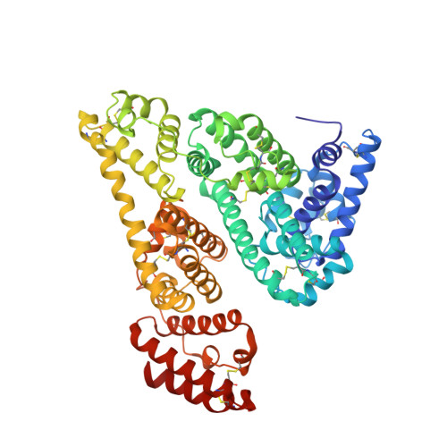
Serum albumin (SA) is the most abundant protein in plasma and is the main transporter of molecules in the circulatory system of all vertebrates, with applications in medicine, the pharmaceutical industry and molecular biology. It is known that albumins from different organisms vary in sequence; thus, it is important to know the impact of the amino-acid sequence on the three-dimensional structure and ligand-binding properties. Here, crystal structures of ovine (OSA) and caprine (CSA) serum albumins, isolated from sheep and goat blood, are described, as well those of their complexes with 3,5-diiodosalicylic acid (DIS): OSA-DIS (2.20 Å resolution) and CSA-DIS (1.78 Å resolution). The ligand-free OSA structure was determined in the trigonal space group P3 2 21 at 2.30 Å resolution, while that of CSA in the orthorhombic space group P2 1 2 1 2 1 was determined at 1.94 Å resolution. Both albumins are also capable of crystallizing in the triclinic space group P1, giving isostructural crystals that diffract to around 2.5 Å resolution. A comparison of OSA and CSA with the closely related bovine serum albumin (BSA) shows both similarities and differences in the distribution of DIS binding sites. The investigated serum albumins from domesticated ruminants in their complexes with DIS are also compared with the analogous structures of equine and human serum albumins (ESA-DIS and HSA-DIS). Surprisingly, despite 98% sequence similarity, OSA binds only two molecules of DIS, whereas CSA binds six molecules of this ligand. Moreover, the binding of DIS to OSA and CSA introduced changes in the overall architecture of the proteins, causing not only different conformations of the amino-acid side chains in the binding pockets, but also a significant shift of the whole helices, changing the volume of the binding cavities.
Organizational Affiliation:
Institute of Technical Biochemistry, Faculty of Biotechnology and Food Sciences, Lodz University of Technology, Stefanowskiego 4/10, 90-924 Lodz, Poland.