5OOI

STRUCTURE OF PROTEIN KINASE CK2 CATALYTIC SUBUNIT (ISOFORM CK2ALPHA') IN COMPLEX WITH THE INDENOINDOLE-TYPE INHIBITOR 4P


Experimental Data Snapshot

  • Method: X-RAY DIFFRACTION
  • Resolution: 2.00 Å
  • R-Value Free: 0.221 
  • R-Value Work: 0.174 
  • R-Value Observed: 0.175 

wwPDB Validation   3D Report Full Report


Ligand Structure Quality Assessment 


This is version 3.1 of the entry. See complete history


Literature

Unexpected Binding Mode of a Potent Indeno[1,2-b]indole-Type Inhibitor of Protein Kinase CK2 Revealed by Complex Structures with the Catalytic Subunit CK2 alpha and Its Paralog CK2 alpha '.

Hochscherf, J.Lindenblatt, D.Witulski, B.Birus, R.Aichele, D.Marminon, C.Bouaziz, Z.Le Borgne, M.Jose, J.Niefind, K.

(2017) Pharmaceuticals (Basel) 10

  • DOI: https://doi.org/10.3390/ph10040098
  • Primary Citation of Related Structures:  
    5OMY, 5ONI, 5OOI

  • PubMed Abstract: 

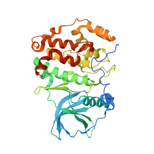
    Protein kinase CK2, a member of the eukaryotic protein kinase superfamily, is associated with cancer and other human pathologies and thus an attractive drug target. The indeno[1,2- b ]indole scaffold is a novel lead structure to develop ATP-competitive CK2 inhibitors. Some indeno[1,2- b ]indole-based CK2 inhibitors additionally obstruct ABCG2, an ABC half transporter overexpressed in breast cancer and co-responsible for drug efflux and resistance. Comprehensive derivatization studies revealed substitutions of the indeno[1,2- b ]indole framework that boost either the CK2 or the ABCG2 selectivity or even support the dual inhibition potential. The best indeno[1,2- b ]indole-based CK2 inhibitor described yet (IC 50 = 25 nM) is 5-isopropyl-4-(3-methylbut-2-enyl-oxy)-5,6,7,8-tetrahydroindeno[1,2- b ]indole-9,10-dione ( 4p ). Herein, we demonstrate the membrane permeability of 4p and describe co-crystal structures of 4p with CK2α and CK2α', the paralogs of human CK2 catalytic subunit. As expected, 4p occupies the narrow, hydrophobic ATP site of CK2α/CK2α', but surprisingly with a unique orientation: its hydrophobic substituents point towards the solvent while its two oxo groups are hydro-gen-bonded to a hidden water molecule. An equivalent water molecule was found in many CK2α structures, but never as a critical mediator of ligand binding. This unexpected binding mode is independent of the interdomain hinge/helix αD region conformation and of the salt content in the crystallization medium.


  • Organizational Affiliation

    Department für Chemie, Institut für Biochemie, Universität zu Köln, Zülpicher Straße 47, D-50674 Köln, Germany. j.hochscherf@uni-koeln.de.


Macromolecules
Find similar proteins by:  (by identity cutoff)  |  3D Structure
Entity ID: 1
MoleculeChains Sequence LengthOrganismDetailsImage
Casein kinase II subunit alpha'
A, B
364Homo sapiensMutation(s): 1 
Gene Names: CSNK2A2CK2A2
EC: 2.7.11.1
UniProt & NIH Common Fund Data Resources
Find proteins for P19784 (Homo sapiens)
Explore P19784 
Go to UniProtKB:  P19784
PHAROS:  P19784
GTEx:  ENSG00000070770 
Entity Groups  
Sequence Clusters30% Identity50% Identity70% Identity90% Identity95% Identity100% Identity
UniProt GroupP19784
Sequence Annotations
Expand
  • Reference Sequence
Small Molecules
Ligands 4 Unique
IDChains Name / Formula / InChI Key2D Diagram3D Interactions
9YE
Query on 9YE

Download Ideal Coordinates CCD File 
C [auth A],
J [auth B]
4-(3-methylbut-2-enoxy)-5-propan-2-yl-7,8-dihydro-6~{H}-indeno[1,2-b]indole-9,10-dione
C23 H25 N O3
IGWUYWFNHIJBBD-UHFFFAOYSA-N
EDO
Query on EDO

Download Ideal Coordinates CCD File 
D [auth A],
E [auth A],
F [auth A]
1,2-ETHANEDIOL
C2 H6 O2
LYCAIKOWRPUZTN-UHFFFAOYSA-N
ACT
Query on ACT

Download Ideal Coordinates CCD File 
H [auth A],
I [auth A],
K [auth B],
L [auth B]
ACETATE ION
C2 H3 O2
QTBSBXVTEAMEQO-UHFFFAOYSA-M
NA
Query on NA

Download Ideal Coordinates CCD File 
G [auth A]SODIUM ION
Na
FKNQFGJONOIPTF-UHFFFAOYSA-N
Experimental Data & Validation

Experimental Data

  • Method: X-RAY DIFFRACTION
  • Resolution: 2.00 Å
  • R-Value Free: 0.221 
  • R-Value Work: 0.174 
  • R-Value Observed: 0.175 
  • Space Group: P 21 21 21
Unit Cell:
Length ( Å )Angle ( ˚ )
a = 46.488α = 90
b = 112.128β = 90
c = 143.692γ = 90
Software Package:
Software NamePurpose
PHENIXrefinement
XDSdata reduction
Aimlessdata scaling
PHASERphasing

Structure Validation

View Full Validation Report


Ligand Structure Quality Assessment 


Entry History & Funding Information

Deposition Data


Funding OrganizationLocationGrant Number
German Research FoundationGermanyNI 643/4-2

Revision History  (Full details and data files)

  • Version 1.0: 2017-12-27
    Type: Initial release
  • Version 2.0: 2018-10-03
    Changes: Advisory, Atomic model, Data collection, Database references, Derived calculations, Polymer sequence, Source and taxonomy, Structure summary
  • Version 3.0: 2018-10-10
    Changes: Advisory, Atomic model, Data collection, Database references, Derived calculations, Polymer sequence, Source and taxonomy, Structure summary
  • Version 3.1: 2024-01-17
    Changes: Data collection, Database references, Refinement description