5OO9

Streptomyces PAC13 (Y55F) with uridine


Experimental Data Snapshot

  • Method: X-RAY DIFFRACTION
  • Resolution: 1.59 Å
  • R-Value Free: 0.200 
  • R-Value Work: 0.160 
  • R-Value Observed: 0.162 

wwPDB Validation   3D Report Full Report


Ligand Structure Quality Assessment 


This is version 1.2 of the entry. See complete history


Literature

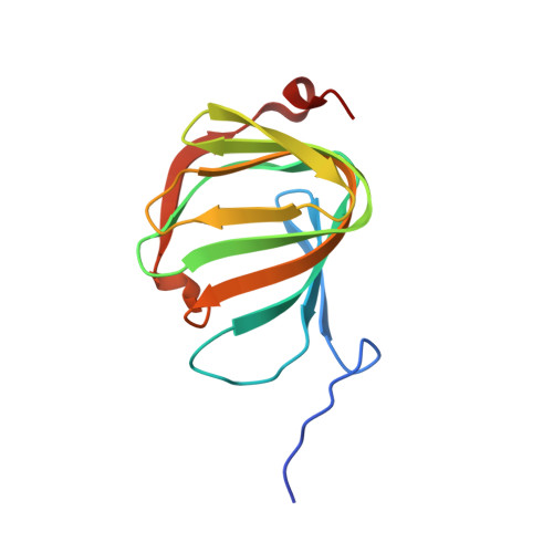
Pac13 is a Small, Monomeric Dehydratase that Mediates the Formation of the 3'-Deoxy Nucleoside of Pacidamycins.

Michailidou, F.Chung, C.W.Brown, M.J.B.Bent, A.F.Naismith, J.H.Leavens, W.J.Lynn, S.M.Sharma, S.V.Goss, R.J.M.

(2017) Angew Chem Int Ed Engl 56: 12492-12497

  • DOI: https://doi.org/10.1002/anie.201705639
  • Primary Citation of Related Structures:  
    5NJN, 5NJO, 5OO4, 5OO5, 5OO8, 5OO9, 5OOA

  • PubMed Abstract: 

    The uridyl peptide antibiotics (UPAs), of which pacidamycin is a member, have a clinically unexploited mode of action and an unusual assembly. Perhaps the most striking feature of these molecules is the biosynthetically unique 3'-deoxyuridine that they share. This moiety is generated by an unusual, small and monomeric dehydratase, Pac13, which catalyses the dehydration of uridine-5'-aldehyde. Here we report the structural characterisation of Pac13 with a series of ligands, and gain insight into the enzyme's mechanism demonstrating that H42 is critical to the enzyme's activity and that the reaction is likely to proceed via an E1cB mechanism. The resemblance of the 3'-deoxy pacidamycin moiety with the synthetic anti-retrovirals, presents a potential opportunity for the utilisation of Pac13 in the biocatalytic generation of antiviral compounds.


  • Organizational Affiliation

    School of Chemistry, University of St Andrews, North Haugh, St Andrews, Fife, KY16 9ST, UK.


Macromolecules
Find similar proteins by:  (by identity cutoff)  |  3D Structure
Entity ID: 1
MoleculeChains Sequence LengthOrganismDetailsImage
Putative cupin_2 domain-containing isomerase124Streptomyces coeruleorubidusMutation(s): 1 
Gene Names: pac13pacM
UniProt
Find proteins for E2EKP5 (Streptomyces coeruleorubidus)
Explore E2EKP5 
Go to UniProtKB:  E2EKP5
Entity Groups  
Sequence Clusters30% Identity50% Identity70% Identity90% Identity95% Identity100% Identity
UniProt GroupE2EKP5
Sequence Annotations
Expand
  • Reference Sequence
Small Molecules
Ligands 1 Unique
IDChains Name / Formula / InChI Key2D Diagram3D Interactions
URI
Query on URI

Download Ideal Coordinates CCD File 
B [auth A]URIDINE
C9 H12 N2 O6
DRTQHJPVMGBUCF-XVFCMESISA-N
Experimental Data & Validation

Experimental Data

  • Method: X-RAY DIFFRACTION
  • Resolution: 1.59 Å
  • R-Value Free: 0.200 
  • R-Value Work: 0.160 
  • R-Value Observed: 0.162 
  • Space Group: P 32 2 1
Unit Cell:
Length ( Å )Angle ( ˚ )
a = 66.604α = 90
b = 66.604β = 90
c = 54.257γ = 120
Software Package:
Software NamePurpose
REFMACrefinement
XDSdata reduction
Aimlessdata scaling

Structure Validation

View Full Validation Report


Ligand Structure Quality Assessment 


Entry History 

Deposition Data

Revision History  (Full details and data files)

  • Version 1.0: 2017-08-23
    Type: Initial release
  • Version 1.1: 2017-09-13
    Changes: Database references
  • Version 1.2: 2017-10-11
    Changes: Database references