5OMT

Endonuclease NucB


Experimental Data Snapshot

  • Method: X-RAY DIFFRACTION
  • Resolution: 1.35 Å
  • R-Value Free: 0.186 
  • R-Value Work: 0.148 
  • R-Value Observed: 0.150 

wwPDB Validation   3D Report Full Report


This is version 1.2 of the entry. See complete history


Literature

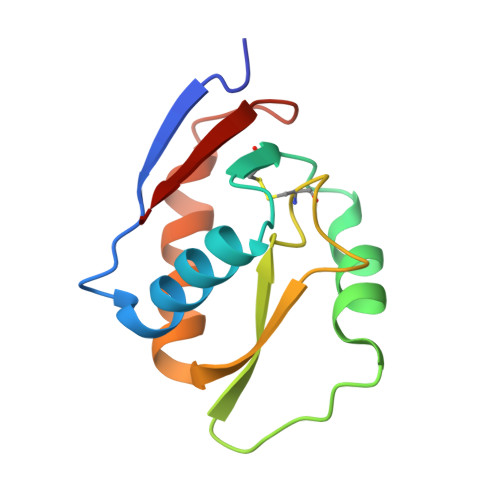
Crystal structure of NucB, a biofilm-degrading endonuclease.

Basle, A.Hewitt, L.Koh, A.Lamb, H.K.Thompson, P.Burgess, J.G.Hall, M.J.Hawkins, A.R.Murray, H.Lewis, R.J.

(2018) Nucleic Acids Res 46: 473-484

  • DOI: https://doi.org/10.1093/nar/gkx1170
  • Primary Citation of Related Structures:  
    5OMT

  • PubMed Abstract: 

    Bacterial biofilms are a complex architecture of cells that grow on moist interfaces, and are held together by a molecular glue of extracellular proteins, sugars and nucleic acids. Biofilms are particularly problematic in human healthcare as they can coat medical implants and are thus a potential source of disease. The enzymatic dispersal of biofilms is increasingly being developed as a new strategy to treat this problem. Here, we have characterized NucB, a biofilm-dispersing nuclease from a marine strain of Bacillus licheniformis, and present its crystal structure together with the biochemistry and a mutational analysis required to confirm its active site. Taken together, these data support the categorization of NucB into a unique subfamily of the ββα metal-dependent non-specific endonucleases. Understanding the structure and function of NucB will facilitate its future development into an anti-biofilm therapeutic agent.


  • Organizational Affiliation

    Institute for Cell and Molecular Biosciences, Faculty of Medical Sciences, Newcastle University, Newcastle upon Tyne NE2 4HH, UK.


Macromolecules
Find similar proteins by:  (by identity cutoff)  |  3D Structure
Entity ID: 1
MoleculeChains Sequence LengthOrganismDetailsImage
NucB110Bacillus licheniformisMutation(s): 0 
Gene Names: nucB
UniProt
Find proteins for F1BV52 (Bacillus licheniformis)
Explore F1BV52 
Go to UniProtKB:  F1BV52
Entity Groups  
Sequence Clusters30% Identity50% Identity70% Identity90% Identity95% Identity100% Identity
UniProt GroupF1BV52
Sequence Annotations
Expand
  • Reference Sequence
Experimental Data & Validation

Experimental Data

  • Method: X-RAY DIFFRACTION
  • Resolution: 1.35 Å
  • R-Value Free: 0.186 
  • R-Value Work: 0.148 
  • R-Value Observed: 0.150 
  • Space Group: P 41 21 2
Unit Cell:
Length ( Å )Angle ( ˚ )
a = 59.955α = 90
b = 59.955β = 90
c = 66.604γ = 90
Software Package:
Software NamePurpose
XDSdata scaling
Aimlessdata scaling
SHELXCDphasing
SHELXEmodel building
ARP/wARPmodel building
BUCCANEERmodel building
REFMACrefinement
Cootmodel building

Structure Validation

View Full Validation Report


Entry History 

Deposition Data

Revision History  (Full details and data files)

  • Version 1.0: 2017-11-15
    Type: Initial release
  • Version 1.1: 2017-12-06
    Changes: Database references
  • Version 1.2: 2018-01-24
    Changes: Database references