5OEG

Molecular tweezers modulate 14-3-3 protein-protein interactions


Experimental Data Snapshot

  • Method: X-RAY DIFFRACTION
  • Resolution: 3.15 Å
  • R-Value Free: 0.266 
  • R-Value Work: 0.181 
  • R-Value Observed: 0.185 

wwPDB Validation   3D Report Full Report


Ligand Structure Quality Assessment 


This is version 2.2 of the entry. See complete history


Literature

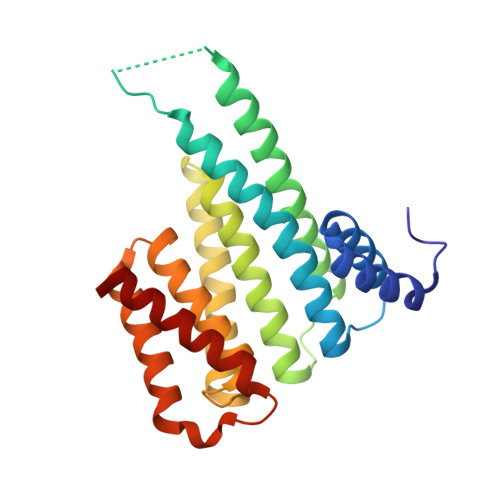
Molecular tweezers modulate 14-3-3 protein-protein interactions.

Bier, D.Rose, R.Bravo-Rodriguez, K.Bartel, M.Ramirez-Anguita, J.M.Dutt, S.Wilch, C.Klarner, F.G.Sanchez-Garcia, E.Schrader, T.Ottmann, C.

(2013) Nat Chem 5: 234-239

  • DOI: https://doi.org/10.1038/nchem.1570
  • Primary Citation of Related Structures:  
    5OEG, 5OEH

  • PubMed Abstract: 

    Supramolecular chemistry has recently emerged as a promising way to modulate protein functions, but devising molecules that will interact with a protein in the desired manner is difficult as many competing interactions exist in a biological environment (with solvents, salts or different sites for the target biomolecule). We now show that lysine-specific molecular tweezers bind to a 14-3-3 adapter protein and modulate its interaction with partner proteins. The tweezers inhibit binding between the 14-3-3 protein and two partner proteins--a phosphorylated (C-Raf) protein and an unphosphorylated one (ExoS)--in a concentration-dependent manner. Protein crystallography shows that this effect arises from the binding of the tweezers to a single surface-exposed lysine (Lys214) of the 14-3-3 protein in the proximity of its central channel, which normally binds the partner proteins. A combination of structural analysis and computer simulations provides rules for the tweezers' binding preferences, thus allowing us to predict their influence on this type of protein-protein interactions.


  • Organizational Affiliation

    Chemical Genomics Centre of the Max-Planck-Society, Otto-Hahn-Strasse 15, 44227 Dortmund, Germany.


Macromolecules
Find similar proteins by:  (by identity cutoff)  |  3D Structure
Entity ID: 1
MoleculeChains Sequence LengthOrganismDetailsImage
14-3-3 protein sigma235Homo sapiensMutation(s): 0 
Gene Names: SFNHME1
UniProt & NIH Common Fund Data Resources
Find proteins for P31947 (Homo sapiens)
Explore P31947 
Go to UniProtKB:  P31947
PHAROS:  P31947
GTEx:  ENSG00000175793 
Entity Groups  
Sequence Clusters30% Identity50% Identity70% Identity90% Identity95% Identity100% Identity
UniProt GroupP31947
Sequence Annotations
Expand
  • Reference Sequence
Small Molecules
Ligands 2 Unique
IDChains Name / Formula / InChI Key2D Diagram3D Interactions
9SZ
Query on 9SZ

Download Ideal Coordinates CCD File 
B [auth A](1R,5S,9S,16R,20R,24S,28S,35R)-3,22-Bis(dihydroxyphosphoryloxy)tridecacyclo[22.14.1.15,20.19,16.128,35.02,23.04,21.06,19.08,17.010,15.025,38.027,36.029,34]dotetraconta-2(23),3,6,8(17),10,12,14,18,21,25,27(36),29,31,33,37-pentadecaene
C42 H32 O8 P2
BZESKQAEKFQSPD-FCODNRGNSA-N
CL
Query on CL

Download Ideal Coordinates CCD File 
C [auth A]CHLORIDE ION
Cl
VEXZGXHMUGYJMC-UHFFFAOYSA-M
Experimental Data & Validation

Experimental Data

  • Method: X-RAY DIFFRACTION
  • Resolution: 3.15 Å
  • R-Value Free: 0.266 
  • R-Value Work: 0.181 
  • R-Value Observed: 0.185 
  • Space Group: C 2 2 2
Unit Cell:
Length ( Å )Angle ( ˚ )
a = 63.5α = 90
b = 154.71β = 90
c = 77.01γ = 90
Software Package:
Software NamePurpose
XSCALEdata scaling
PHASERphasing
REFMACrefinement
PDB_EXTRACTdata extraction
XDSdata reduction

Structure Validation

View Full Validation Report


Ligand Structure Quality Assessment 


Entry History 

Deposition Data

Revision History  (Full details and data files)

  • Version 1.0: 2017-07-26
    Type: Initial release
  • Version 2.0: 2020-04-29
    Changes: Atomic model, Derived calculations
  • Version 2.1: 2024-01-10
    Changes: Data collection, Database references, Derived calculations, Structure summary
  • Version 2.2: 2024-01-17
    Changes: Refinement description