5OAT

PINK1 structure


Experimental Data Snapshot

  • Method: X-RAY DIFFRACTION
  • Resolution: 2.78 Å
  • R-Value Free: 0.246 
  • R-Value Work: 0.205 
  • R-Value Observed: 0.207 

wwPDB Validation   3D Report Full Report


This is version 1.1 of the entry. See complete history


Literature

Structure of PINK1 and mechanisms of Parkinson's disease associated mutations.

Kumar, A.Tamjar, J.Waddell, A.D.Woodroof, H.I.Raimi, O.G.Shaw, A.M.Peggie, M.Muqit, M.M.van Aalten, D.M.

(2017) Elife 6

  • DOI: https://doi.org/10.7554/eLife.29985
  • Primary Citation of Related Structures:  
    5OAT

  • PubMed Abstract: 

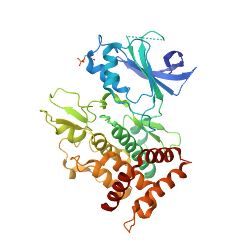
    Mutations in the human kinase PINK1 (hPINK1) are associated with autosomal recessive early-onset Parkinson's disease (PD). hPINK1 activates Parkin E3 ligase activity, involving phosphorylation of ubiquitin and the Parkin ubiquitin-like (Ubl) domain via as yet poorly understood mechanisms. hPINK1 is unusual amongst kinases due to the presence of three loop insertions of unknown function. We report the structure of Tribolium castaneum PINK1 ( Tc PINK1), revealing several unique extensions to the canonical protein kinase fold. The third insertion, together with autophosphorylation at residue Ser205, contributes to formation of a bowl-shaped binding site for ubiquitin. We also define a novel structural element within the second insertion that is held together by a distal loop that is critical for Tc PINK1 activity. The structure of Tc PINK1 explains how PD-linked mutations that lie within the kinase domain result in hPINK1 loss-of-function and provides a platform for the exploration of small molecule modulators of hPINK1.


  • Organizational Affiliation

    Division of Gene Regulation and Expression, University of Dundee, Dundee, United Kingdom.


Macromolecules
Find similar proteins by:  (by identity cutoff)  |  3D Structure
Entity ID: 1
MoleculeChains Sequence LengthOrganismDetailsImage
Serine/threonine-protein kinase PINK1, mitochondrial-like Protein
A, B, C, D, E
A, B, C, D, E, F
411Tribolium castaneumMutation(s): 0 
Gene Names: TcasGA2_TC013202
UniProt
Find proteins for D6WMX4 (Tribolium castaneum)
Explore D6WMX4 
Go to UniProtKB:  D6WMX4
Entity Groups  
Sequence Clusters30% Identity50% Identity70% Identity90% Identity95% Identity100% Identity
UniProt GroupD6WMX4
Sequence Annotations
Expand
  • Reference Sequence
Experimental Data & Validation

Experimental Data

  • Method: X-RAY DIFFRACTION
  • Resolution: 2.78 Å
  • R-Value Free: 0.246 
  • R-Value Work: 0.205 
  • R-Value Observed: 0.207 
  • Space Group: P 1 21 1
Unit Cell:
Length ( Å )Angle ( ˚ )
a = 84.915α = 90
b = 116.736β = 94.29
c = 179.344γ = 90
Software Package:
Software NamePurpose
REFMACrefinement
XDSdata reduction
Aimlessdata scaling
CRANK2phasing

Structure Validation

View Full Validation Report


Entry History & Funding Information

Deposition Data


Funding OrganizationLocationGrant Number
Wellcome TrustUnited Kingdom101022/Z/13/Z
Wellcome TrustUnited Kingdom110061

Revision History  (Full details and data files)

  • Version 1.0: 2017-10-11
    Type: Initial release
  • Version 1.1: 2017-10-18
    Changes: Database references